This content is also available in:
![]() Italiano
Italiano ![]() Português
Português ![]() Deutsch
Deutsch ![]() Čeština
Čeština ![]() Română
Română ![]() Türkçe
Türkçe
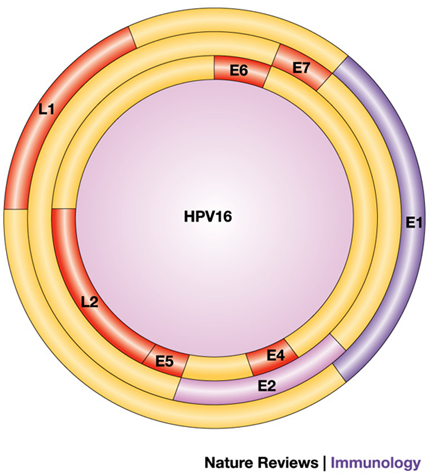
The key proteins coded by HPV DNA
HPV DNA is 6800-8000 base pairs long, which when translated, codes for 8 Genes. The genes are broken down in to early and late phase, indicating which stage of basal cell infection they are introduced. E1, E2, E4, E5, E6, and E7 make up the early phase genes, L1 and L2 are the late phase genes. The key proteins are shown in Figure 3.6.
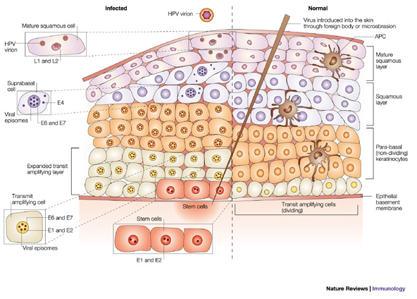
Mechanism of HPV infection
As previously mentioned, the squamous epithelium acts as a protective skin to keep pathogens from infecting the underlying tissue. Even though the cervix is not an externally exposed tissue it is still subject to the external environment, especially during sexual intercourse. The HPV is able to get through the protective layers of the squamous epithelium through epithelial abrasions to commence its infective life cycle.
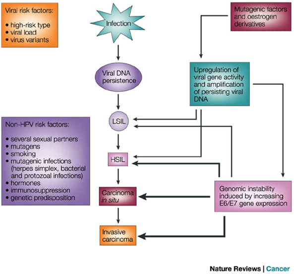
HPV integration into the host genome
There are two main outcomes from the integration of viral DNA into the host genome that can eventually lead to tumour formation.
- Blocking the cells apoptotic pathway
- Blocking synthesis regulatory proteins leading to uncontrolled mitosis
There are two integral genes in the HPV genome that play a central role in tumour formation, E6, which inhibits the role of p53 and E7, which inhibits the role of the retinoblastoma protein (Rb). In normal physiology, p53 and Rb are involved in the regulation of cells with damaged/mutated DNA.
The p53 gene codes for a protein that is vital in protecting us from cells with damaged DNA. In health, the p53 protein binds to damaged DNA and becomes active. Activation of p53 leads to the attraction of other proteins, which ultimately leads to apoptosis (programmed cell death) of the cell that has damaged DNA. When E6 protein of HPV binds to the host p53 protein, it causes a structural change and the variant of p53 is no longer able to bind to damaged DNA. As a consequence of this, the apoptosis cascade involving p21 is unavailable to act as the ‘stop signal’ in mitosis so the mutated cell is able to divide uncontrollably.
The Rb gene codes for a protein that restricts the cells ability to replicate: Rb prevents inhibiting progression from the gap phase to the synthesis phase of the G1 mytotic cycle. Simply put, when the E7 protein binds to and degrades Rb protein, it is no longer functional and cell proliferation is left unchecked.
Learning points from Chapter 3
|