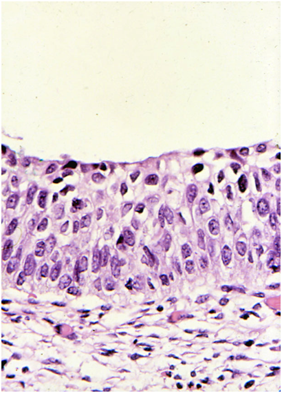
Cytological features of high-grade dyskaryosis (moderate) – HSIL favour CIN2
Presentation
The cells are usually seen in sheets or as separated cells with more cytoplasm than usually seen in CIN3. Crowded overlapping clusters of cells are less likely to be reported as moderate dyskaryosis because the NC ratio in such cells may be difficult to assess.
Nuclei
The nuclear features of dyskaryosis are similar to those described above for HSIL but may be less pronounced. The nuclei may be hyperchromatic, normochromic or hypochromatic.
Cytoplasm
The cytoplasm will usually be similar to that of an intermediate cell but the diameter smaller. Cells with more maturation, suggesting LSIL, may be present alongside HSIL – indicating either surface maturation of the cells or coexisting LSIL: see Figure 9c-7 (a-b).
Nuclear/cytoplasmic ratio
Moderate dyskaryosis is usually reported when the NC ratio is approximately 50% in most of the cells.
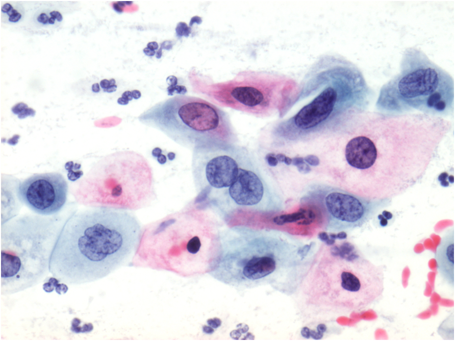
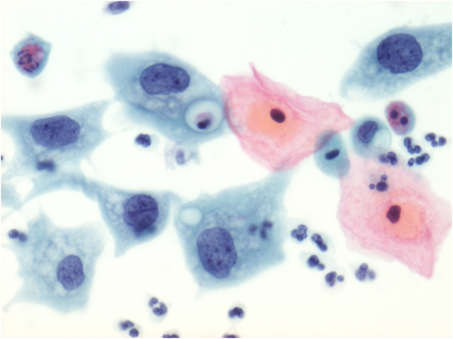
Figure 9c-7 (a-c). High-grade dyskaryosis (moderate) confirmed on histology to be CIN2
(a) Small sheets of cells with nuclear features of dyskaryosis and cytoplasm varying between approximately 50% and more than 50%. Some surface maturation is present accounting for the cells with a higher NC ratio. The nuclei are slightly hyperchromatic.
Distinction between HSIL (favour CIN2) and LSIL
The critical distinction is between HSIL and LSIL. As both may be present on the same slide, rather more frequently with CIN2 than CIN3, and CIN2 is characterised by surface maturation this may sometimes be difficult to decide. A judgement must be made after examining the whole slide based on the degree of nuclear abnormality as well as the predominant NC ratio. The most severe abnormality should be reported.
Furthermore, care must be taken to avoid over-diagnosis of immature squamous metaplasia or atrophic epithelium as HSIL favour CIN2 (moderate dyskaryosis): see below in this chapter in the section on ASC-US
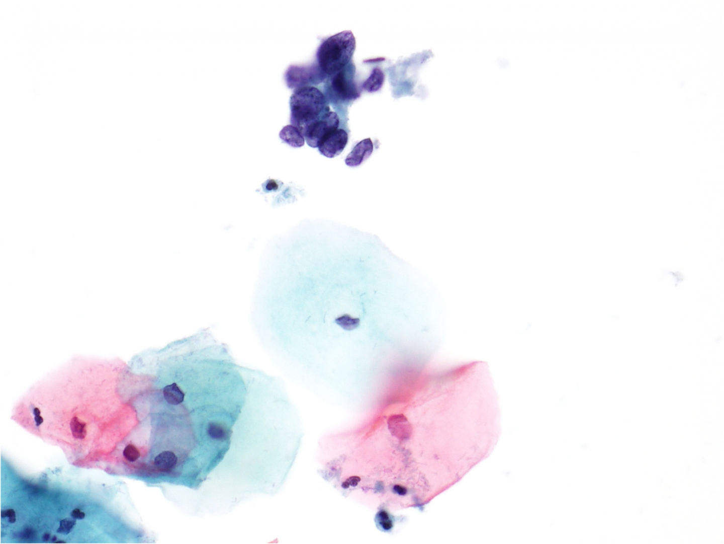
The difficulty in distinguishing certain cases of HSIL from LSIL (in terms of moderate versus mild dyskaryosis) is demonstrated by responses received for two images from a website questionnaire circulated before the BSCC Terminology Conference in 2002 (Figure 9c-8 a-b). The majority correctly assessed the images as high-grade (a) and low-grade (b) respectively but there was considerable observer variation between mild and moderate dyskaryosis in both cases. It should be noted that the nuclei are larger in LSIL than HSIL although the NC ratio is greater in the latter.
The important thing with both these cases is the presence of irregular chromatin distribution and nuclear membranes (dyskaryosis) allowing a definitive diagnosis of SIL to be made rather than atypical squamous cells of undetermined significance (ASC-US) or cannot exclude HSIL (ASC-H). On rare occasions such samples may be reported as dyskaryosis of uncertain grade (Denton et al. 2008) and managed as for high-grade (moderate).
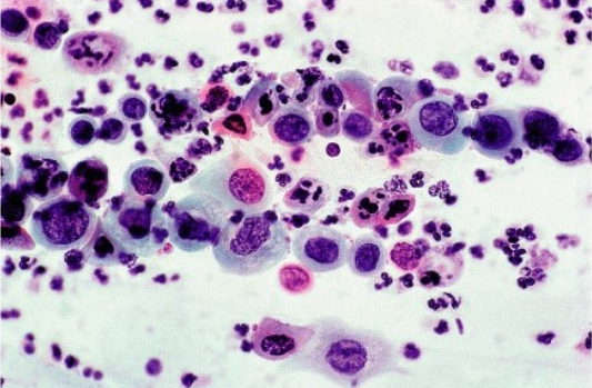
Figure 9c-8 (a-b). Two images of SIL from the BSCC Terminology Conference 2002.
High-grade (moderate) dyskaryosis (CIN2 on histology). Cells with a high NC ratio are intermingled with more mature cells. BSCC respondent assessments: 19% severe, 52% moderate, 27% mild, 2% borderline
| Distinction between HSIL and LSILNuclei tend to be larger in LSIL than HSILNC is increased in HSIL largely because of reduced cytoplasmic sizeCytoplasmic maturation may be seen in some cells in HSIL favour CIN2 (LSIL may coexist)Dyskaryosis is seen in HSIL and LSILIf in doubt report as SIL of uncertain grade rather than ASC-H |
Cytological features of severe dyskaryosis (HSIL favour CIN3)
Presentation
The cells will appear on the cytology preparation both single cells and, frequently, as dense hyperchromatic sheets and clusters. Generally speaking there will be abundant material on the slide but that is not always the case. A prominent feature is that there will be single cells dissociating from the crowded groups and seen as isolated cells between the cell groups. A neutrophil polymorph exudate is often present.
Nucleus
The nucleus is usually (but not always) hyperchromatic, with coarse, irregularly distributed, clumped chromatin. The nucleus shows irregular nuclear borders with a focally thickened nuclear membrane. The NC ratio may approach 100% with only a small amount of cytoplasm remaining. The cell size be only slightly enlarged and the diagnosis depends on the nuclear features and NC ratio.
Cytoplasm
In most instances the cytoplasmic diameter is less than a parabasal or immature metaplastic cell; it may be inconspicuous, particularly in hyperchromatic crowded groups of cells, and may be absent or cytolysed. In some cases the cytoplasm may be more abundant and either keratinising or non-keratinising. Concurrent LSIL and/or koilocytosis may also be present on the slide but this is less frequent than with high-grade dyskaryosis (moderate).
Nuclear/cytoplasmic ratio
This tends to be higher than described in moderate dyskaryosis and is usually uniformly greater than 50% and may approach 100%.
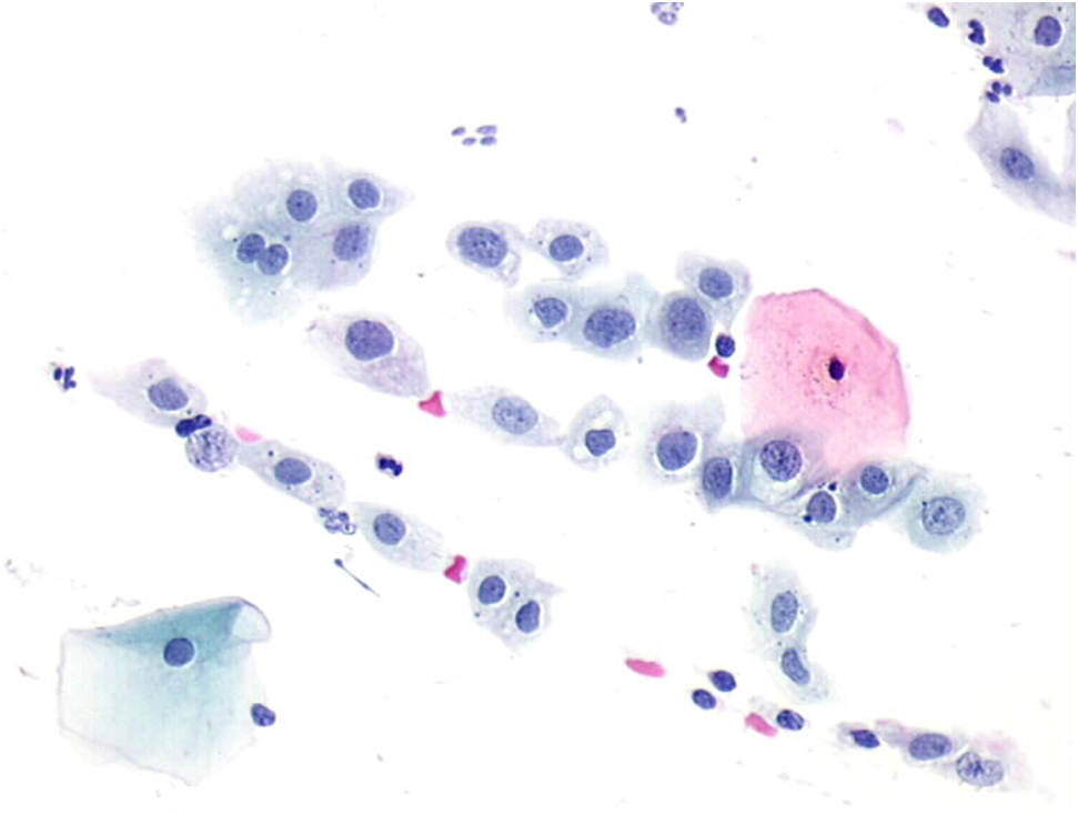
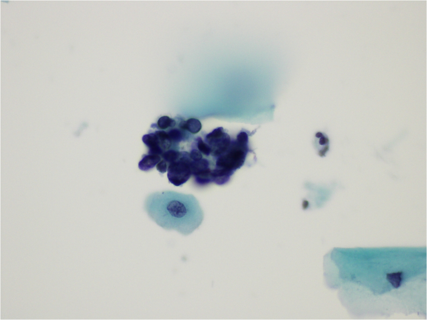
Figure 9c-9 (a-b). High-grade dyskaryosis (severe) in separated cells confirmed on histology to be CIN3
(a) Irregular nuclear membranes and chromatin pattern in hyperchromatic nuclei in a background of inflammation. The NC ratio is well over 50% in most of the cells.
Hyperchromatic crowded cell groups
Crowded cohesive severely dyskaryotic cells are more often seen in CIN3 than CIN2 because they are characterised by scant cytoplasm and a high NC ratio. Examples are shown in Figure 9c-10 below. Nuclear features of HSIL can be seen in separated cells at the margins of the cell groups. Hyperchromatic crowded cell groups may be overlooked on low-power examination, which is a known pitfall in diagnosis.
In some instances hyperchromatic crowded cell groups of HSIL may mimic glandular neoplasia (Figure 9c-11), with which it may coexist. In such cases typical features of CGIN or adenocarcinoma should be looked for elsewhere on the sample.
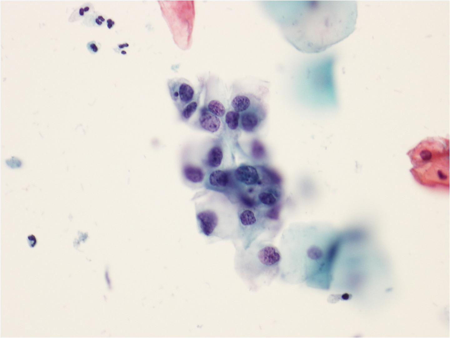
Figure 9c-10 (a-c) Hyperchromatic crowded cell groups – HSIL favour CIN3
(a) Hyperchromatic crowded group of cells, with hardly any associated cytoplasm. There is nuclear size and shape variation with some cells dissociating from the group.
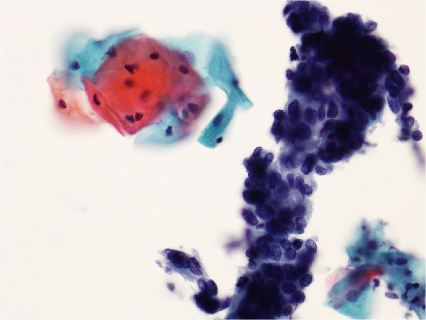
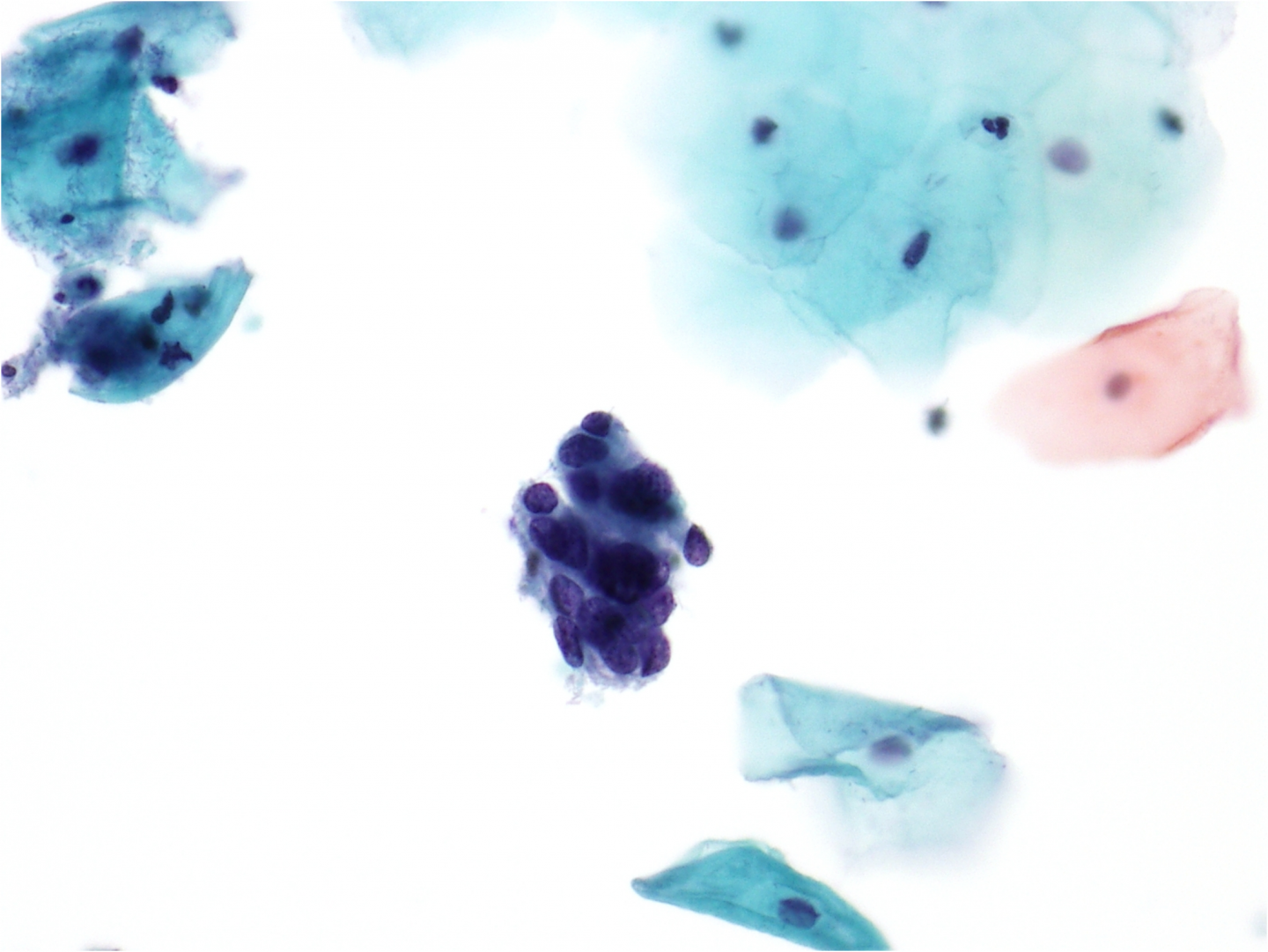
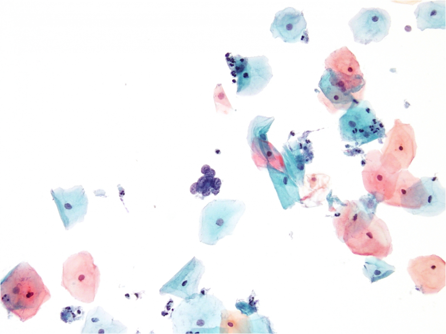
Figure 9c-11 (a-b). Hyperchromatic crowded groups of cell mimicking CGIN
Cohesive clusters of severely dyskaryotic cells with polarisation of mostly round nuclei forming partial rosette-like structures. There were no other features to suggest CGIN and the diagnosis was CIN3.
Differential diagnosis of hyperchromatic crowded cell groups
As the NC ratio tends to be high, the differential diagnosis is unlikely to include LSIL or HSIL favour CIN2 or high-grade dyskaryosis (moderate). The differential diagnosis includes benign entities such as endometrial cells, tubo-endometrioid metaplasia and reactive endocervical cells; and CGIN This type of HSIL may sometimes be classified as ASC-H (see below).