Principles of Immunocytochemistry
There are very many methods for performing immunocytochemistry comprehensive accounts are available on the internet (eg under Immunocytochemical Methods). One well established and useful method will be described here the indirect (two-layer) method using a peroxidase polymer-labelled second antibody. The antigen of interest is preserved in the cells by fixation with alcohol and/or […]
Antibody dilution
Diluent A standard antibody diluent for primary antibodies is 0.01M phosphate-buffered normal saline (0.87% NaCl), pH 7.2 to 7.4 (PBS) containing 0.1% bovine serum albumin (BSA) and 0.1% sodium azide as a preservative. Tris/HCl-buffered saline, pH 7.6 (TBS), may also be used. The BSA acts as an inert protein that can bind to any sticky […]
Controls
A positive control preparation for every antigen should be included in every test run. This will be evidence that the various layers were applied in the correct order and that all the solutions were functional. Any diversion from the normal appearance for the positive control should be an alert that something may be wrong. Ideally, […]
Apparatus
Today many laboratories use automated immunostainers for routine diagnostic immunocytochemistry. Following appropriate fixation (and removal of the PEG layer, if used, in two changes of 95% methanol) the slides are laid in slots in the machine and the preparations are subjected to the various steps in a pre-programmed sequence. Antibodies and their dilution (if not […]
Special considerations for immmunocytopathology
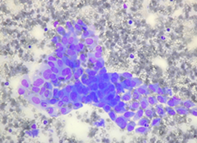
In contrast to histopathological sections, cytological preparations have no tissue context. The initial screening for abnormal cells is therefore essential, and immunocytochemistry will be used to resolve differential diagnostic problems, again relying on the morphology of the cells as well as the antigen localization. Normal cells in sample It is important to remember that some […]
Appendix
1) Espostis fluid Mix together: 100 ml distilled water 45 ml methanol 10 ml glacial acetic acid Store at ambient temperature 2) Buffers 0.01M phosphate-buffered saline (PBS), pH 7.2 -7.4. This buffer can be made from purchased tablets (ensure the molarity and pH are correct) or more cheaply made in the laboratory. Reagents Sodium […]
References
Burry RW. Controls for Immunocytochemistry. An Update. J Histochem Cytochem ( 2011) 59(1): 612. doi: 10.1369/jhc.2010.956920 Kirbis,I.S, Maxwell,P., Flezar,M.S., Miller,K., Ibrahim,M. External quality control for immunocytochemistry on cytology samples: a review of UK NEQAS ICC (cytology module) results. Cytopathology (2011) 22: 230-237. Leong, A., Suthipintawong,C., Vinyuvat,S. Immunostaining of cytologic preparations: a review of technical problems. Appl. Immunohistochem. Mol. […]
Immunocytochemistry
Author: Susan Van Noorden Preservation of Cytological Samples for Immunocytochemistry Foreword Immunocytochemistry is now essential for full and accurate pathological diagnosis of tissue samples whether of formalin-fixed blocks or cell preparations from brushings, scrapings, smears or fine needle aspirates. The success of the method depends to a large extent on the morphological preservation of the […]
Preparation and fixation of samples
If the sample contains enough material, after smears and cytospins have been made for immediate evaluation, the method of choice is to spin down the rest into a cell button which is fixed in 10% formalin (formal saline or phosphate-buffered formalin) followed by dehydration through graded alcohols and paraffin embedding for sectioning. This has the […]