This content is also available in:
![]() Čeština
Čeština
In contrast to histopathological sections, cytological preparations have no tissue context. The initial screening for abnormal cells is therefore essential, and immunocytochemistry will be used to resolve differential diagnostic problems, again relying on the morphology of the cells as well as the antigen localization.
Normal cells in sample
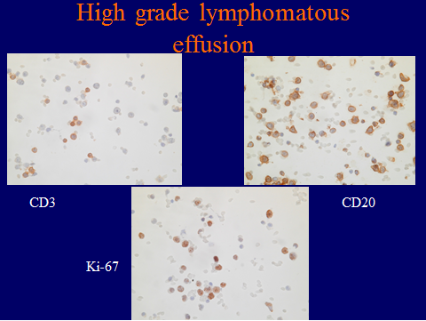
It is important to remember that some immunoreactive cells in a sample may be normal cells and not malignant, for example, CD3-positive T-lymphocytes in a high grade lymphomatous effusion where the CD-20 B-lymphocytes are the malignant cells.
Few abnormal cells in sample
Another problem relates to sampling error and paucity of suspicious cells in the FNA sample. If very few cells show morphological alterations, a single abnormal cell showing positive immunostaining may be enough for diagnosis, but a negative result, in the absence of abnormal cells, would be inadequate. It would be necessary in this case to try the immunostaining on several preparations from the same sample, if possible, or in the last resort to de-stain the Papanicolau stain (after photographing the preparation) and re-stain with immunocytochemistry.
There are a few other resources that may be used if abnormal cells are scarce. If the cell preparation is large enough, such as an LBC sample, it can be divided into two or more parts by drawing a line across the preparation with an isolating pen. Different primary antibodies can be applied to each section of the preparation. Before doing this, stain the preparation lightly with haematoxylin to check that abnormal cells are present in each part. If the expected antigen has a nuclear localization, the light haematoxylin stain can be removed in acid-alcohol before the immunostain is applied. If it is cytoplasmic, this will not be necessary. This method is also applicable to smears, but cytospin preparations would be too small.
Another remedy would be to remove the coverslip from the negative control preparation (provided that it was negative), remove the nuclear stain with acid-alcohol, if necessary, and re-stain with a different primary antibody.
Staining for two antibodies simultaneously in the same sample is possible by a variety of means beyond the scope of this chapter (ref Polak & Van Noorden book or Van der Loos article).
Background staining
If the original sample is very proteinaceous or bloody, background staining may interfere with the final picture, particularly in smears directly from the FNA. The best remedy is to wash the sample in before making the smear or cytospin in a commercial solution such as Cytolite or in Espostis fluid (Appendix) or similar. Post- fixation in formalin may help to lyse red blood cells and blocking of endogenous peroxidase is essential.