There are very many methods for performing immunocytochemistry comprehensive accounts are available on the internet (eg under Immunocytochemical Methods). One well established and useful method will be described here the indirect (two-layer) method using a peroxidase polymer-labelled second antibody.
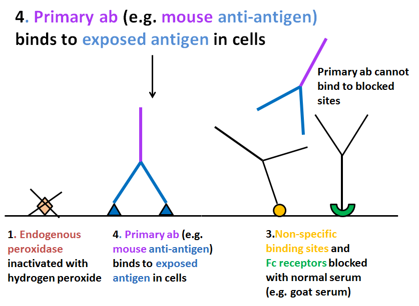
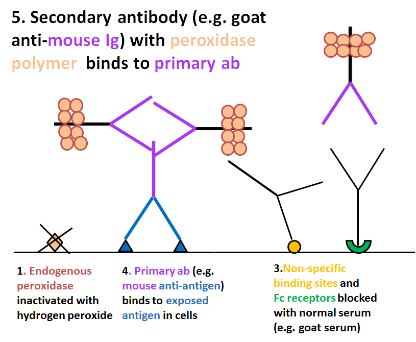
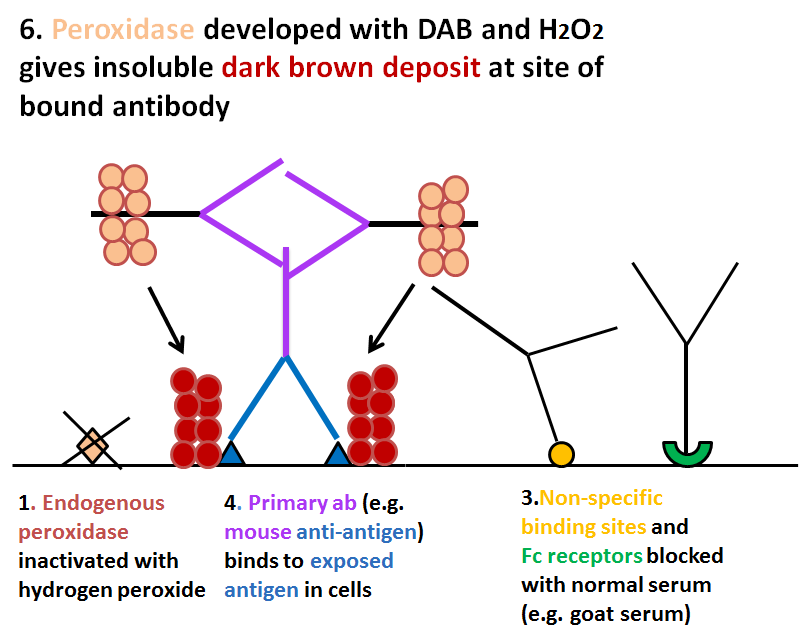
The antigen of interest is preserved in the cells by fixation with alcohol and/or formalin. The primary antibody, usually raised in a rabbit or a mouse, binds to the antigen and excess is washed off with buffer. The secondary antibody is raised (in goat, or other species) to immunoglobulins of the primary antibody host (eg. goat anti-mouse Ig) and binds to the primary antibody at the site where it is attached to its antigen. The secondary antibody is labelled so that the reaction site can be visualised in the microscope. The most efficient label is polymerised horseradish peroxidase which provides a large amount of enzyme label on the second antibody molecule. Following further washing, the peroxidase is reacted with its substrate, hydrogen peroxide, and a chromogen, usually diaminobenzidine (DAB) which gives a dark brown, insoluble precipitate at the site of reaction. The preparation can then be counterstained with haematoxylin, dehydrated in graded alcohols, cleared in a solvent such as xylene and permanently mounted.
Antigen retrieval
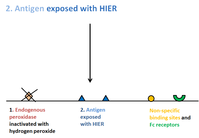
Immunoreactions are often enhanced by heat-induced antigen retrieval (HIER). This is not always necessary for cell preparations that have been fixed in alcohol, but is usually advantageous in revealing the maximum immunoreactivity of fixed antigens, particularly after formalin fixation. HIER is carried out either before or after the peroxidase blocking step. Preparations are immersed in a buffer and heated in a closed plastic container in a microwave oven (domestic oven at full power) or in a pressure cooker or autoclave, for a pre-established time. The period of heating (usually 10 to 20 minutes) will depend on whether the preparations are put into cold buffer and then heated or put directly into hot buffer, and whether they are left to cool in the buffer or are cooled rapidly by running cold water into the container. It is important that they are not lifted out of hot buffer which would immediately evaporate and cause the preparations to become dry. During the heating period in a microwave oven it may be necessary to top up the fluid level at intervals with hot distilled water to replace that which has evaporated and keep the slides immersed. When a convenient retrieval method has been established, this should become standard for the laboratory. The buffer to use should be tested for each new antibody (or use that recommended by the supplier). Some antigens are best revealed after heating in a slightly acidic buffer (0.01M citrate buffer, pH 6.0), others in an alkaline buffer (TRIS/EDTA, pH9.0). These can be easily prepared in the laboratory. Most antibody companies supply appropriate buffers. (For review, see Shi et al, 2011)
Blocking steps
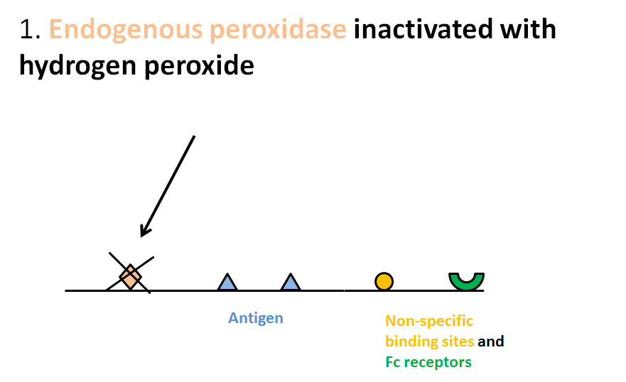
Endogenous peroxidase
Some cells contain peroxidase-like enzymes that must be blocked to prevent them reacting in the final peroxidase label visualisation step, which might confuse the resulting image. Blocking is done before antibodies are applied, using excess of the enzyme substrate, hydrogen peroxide, at a much higher concentration than is used to reveal the antibody-bound enzyme at the end. This effectively prevents further action of the endogenous enzyme.
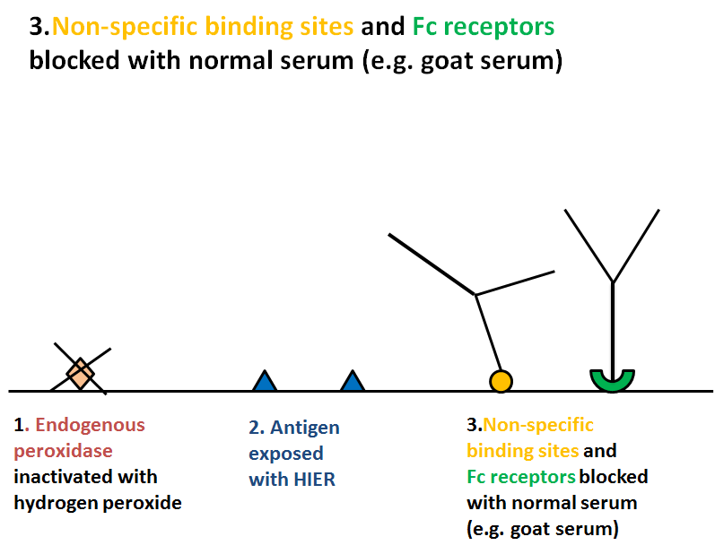
Non-specific background staining
Sometimes Fc receptors on the cells can bind applied antibodies, or proteins in the preparations can react non-specifically with them by charged or hydrophobic reactions. Such non-specifically bound primary or secondary antibodies will subsequently react in the same way as specific ones and give a confusing result. These reactions will be visible in negative control preparations in which the primary antibody is omitted or replaced with an irrelevant antibody. Non-specific reactions of this nature can be blocked by a layer of non-immune serum (from the same species as the secondary antibody) before application of the primary antibody. When the non-specific combining sites are occupied by normal immunoglobulin, the first antibody cannot bind to them and provided the blocking antibody was from the same species as the second antibody that, too, cannot bind.