There is no requirement in TBS to sub-classify HSIL as ‘favour CIN2 or favour CIN3’, or high-grade dyskaryosis as moderate or severe dyskaryosis in the UK system (Denton et al. 2008). The most important distinction to be made on cytology is between LSIL and HSIL.
Nevertheless, the UK system and some others in Europe prefer to diagnose moderate and severe dyskaryosis or dysplasia as such (Herbert et al. 2007) while defining and managing them as high-grade. The advantages of this sub-classification are that i) the differential diagnosis and ii) the positive predictive value (Blanks & Kelly 2010) tend to be different for these entities. This may be important when considering the risk of progression versus likelihood of regression alongside the histological diagnosis when deciding between treatment and surveillance.
Cytological features of HSIL
Presentation
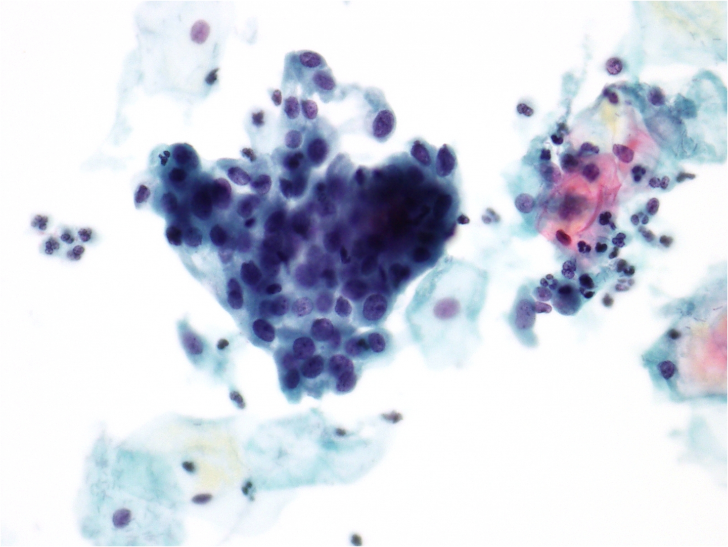
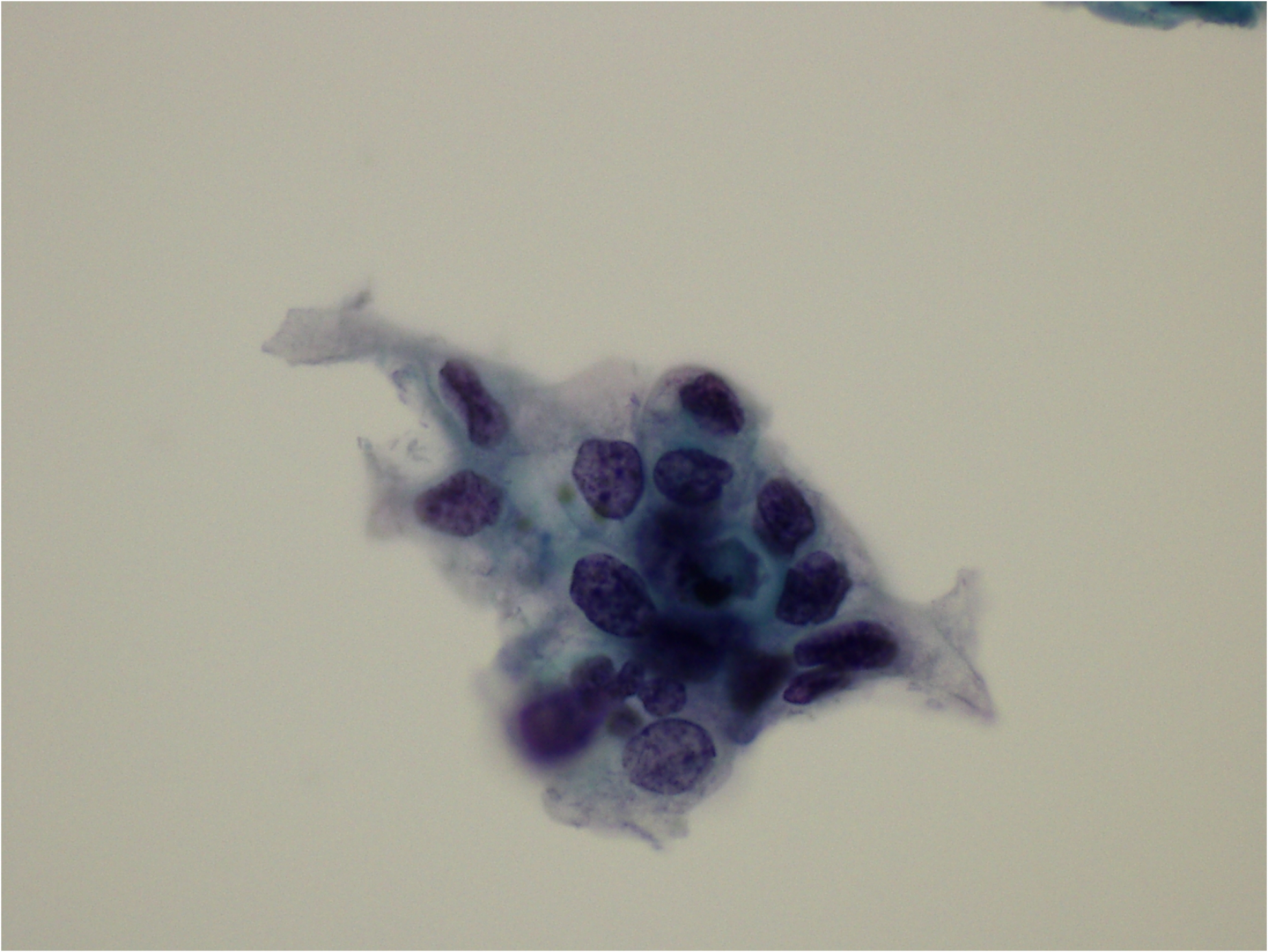
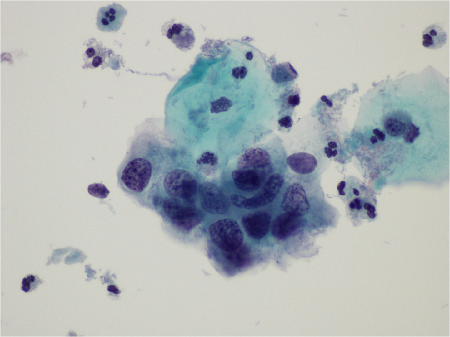
The cells will appear on the cervical smear or LBC preparation as single cells, sheets or crowded groups of cells: where the latter are seen there are usually separated cells at the margins or between the cell groups in which the features of SIL can more clearly be seen. The dyskaryotic cells will be immature indicating incomplete epithelial maturation.
Nucleus
The chromatin is more granular and irregularly distributed than in LSIL. We begin to see true nuclear membrane irregularities with irregular borders and focal thickening, nuclear notches and folds. Where the abnormal cells are shed in sheets, there may be a loss of polarity and some crowding. The nucleus is most often hyperchromatic but may be normochromatic or hypochromatic. The nuclei in HSIL are not necessarily enlarged and may be little larger than that of a normal intermediate cell (Smith & Turnbull 1997; Denton et al. 2008; Wilbur et al. 2015).
Cytoplasm
The cytoplasm will vary in size between rather smaller than a normal intermediate cell and almost complete absence. The amount of cytoplasm is a cardinal feature of the distinction between moderate and severe dyskaryosis (i.e. CIN2 and CIN3).
Nuclear/cytoplasmic ratio
As the cells are less mature than in LSIL there is a higher NC ratio: the nucleus in HSIL occupies at least 50% of the diameter of the cell. The increased NC ratio is more due to reduced cytoplasmic size than nuclear enlargement (Nayer et al. 2004; Slater et al. 2005a, 2005b).
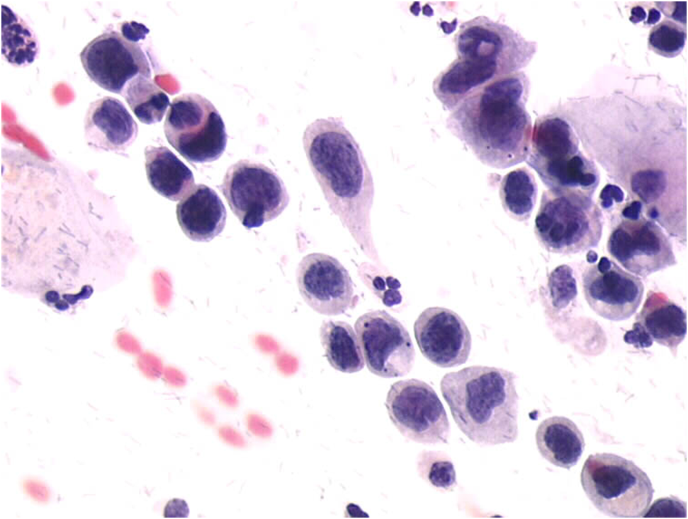
9c-6 (a-d). HSIL – high-grade dyskaryosis
(a) Separated cells in a conventional smear showing a streak of separated cells showing the typical features of high-grade dyskaryosis: irregularly dispersed chromatin and nuclear outlines, immature cytoplasm; NC ratio variable but well over 50% in most of the cells. In this case there is hyperchromasia and the nuclei are slightly enlarged.