This content is also available in:
![]() Italiano
Italiano ![]() Português
Português ![]() Deutsch
Deutsch ![]() Čeština
Čeština ![]() Română
Română ![]() Türkçe
Türkçe
The uterine cervix primarily acts as a physical barrier between the external environment (vaginal canal) and the uterus. The cells lining the endocervical canal produce both acid and neutral mucin, which contains electrolytes (mostly sodium chloride) and simple sugars (glycogen) in colloidal solution. This mucus forms a plug in the os blocking the passage of foreign pathogens/objects from the vaginal vault into the uterus. Immunoglobulins, enzymes, leucocytes and exfoliated squamous epithelial cells can also be found trapped in this mucus, further strengthening the plug and adding bactericidal properties to the mucus.
The mucus plug is not a permanent fixture and hormones determine its consistency. At the time of ovulation, the mucus becomes much thinner and is composed of a fine acellular network of filaments, which facilitates the passage of sperm from the vagina into the uterus and eventually into the fallopian tubes, where fertilisation can take place. During pregnancy the mucus plug becomes thick and protects the developing foetus by sealing the endocervical canal once again.
The cervix and uterus are pliable structures, which contain elastic fibres in addition to muscle. This is most evident during childbirth when the cervix is required to dilate to approximately ten centimetres in diameter to accommodate the passage of the foetus into the vaginal canal.
If fertilisation does not take place; the myometrium of the cervix dilates to allow the passage of menses (broken down lining of the endometrium). This dilatation is known to cause sensations of pain and discomfort commonly referred to as period pain. These physiological features of the cervix are all controlled by the hormones oestrogen and progesterone.
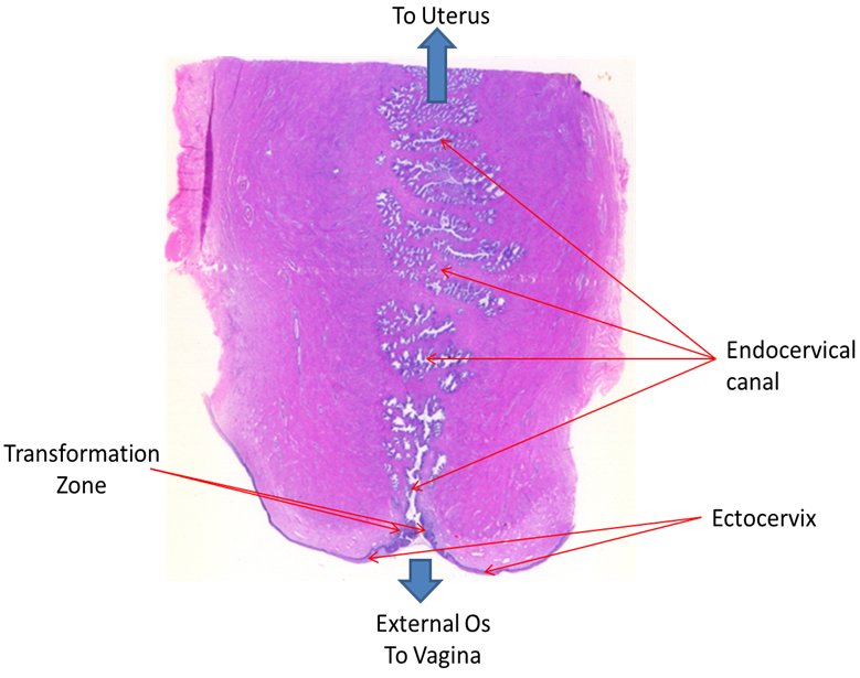
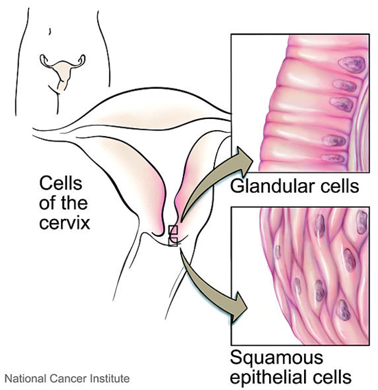
There are two types of epithelium lining the cervix; stratified squamous epithelium on the ectocervix and simple columnar (glandular) epithelium lining the endocervix and crypts.