Clinical features
- Benign tumour/tumour like malformation
- Rare in children
- Association with other malformations and tumours
- X-ray-
- Peripheral coin lesion with well demarcated borders
- Calcification (curvilinear popcorn)-diagnostic-best seen in computed tomography
Fig 88 – Lung Hamartoma Smear from a transthoracic guided fine needle aspiration, Fragment of mesenchyma in a myxoid matrix. (H&E)
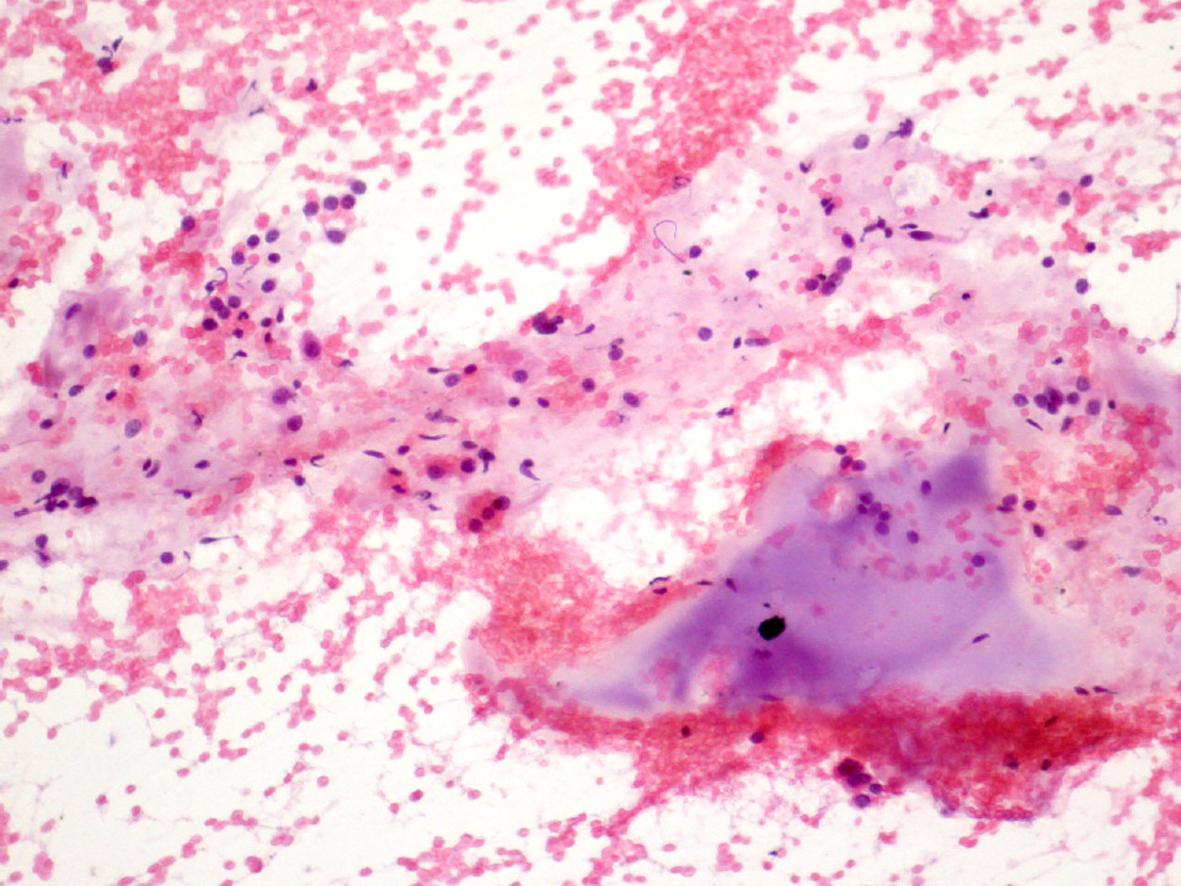
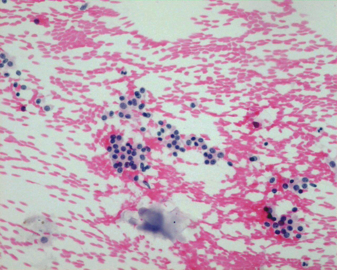
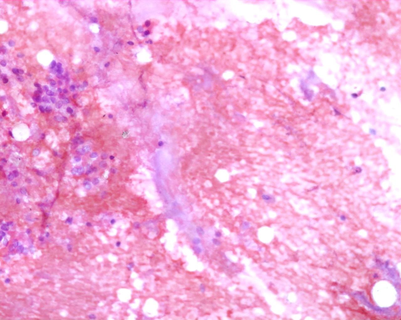
- Malformed cartilage -the presence of cartilage by itself is not enough
- Mesenchymal myxoid matrix (essential to diagnose) (characteristic linear fibrillary parallelism)
- Sheets of bronquial epithelium
Immunocytochemistry
No contributory
Modern Techniques of Diagnosis
No contributory
Differential Diagnosis
- Normal tissue from chest wall or bronchus
- Teratoma (Dermoid)
- More frequent in the mediastinum
- Cartilage can also be seen as well as mesenchymal tissue
- Blastoma
- Cartilage is immature