Clinical features
- Rare before 12 years
- Slight female predominance
- Haemoptysis
- Rarely associated with endocrine symptoms
- Occasionally associated with other endocrine tumours
- Main lobar or segmental bronchi
- In children they are rarely peripheral
Fig 93 – Carcinoid – Smear from a transthoracic guided fine needle aspiration. Uniform population of single round/ oval regular neoplastic cells. Occasionally cells get together in small acini. Clear background (H&E).
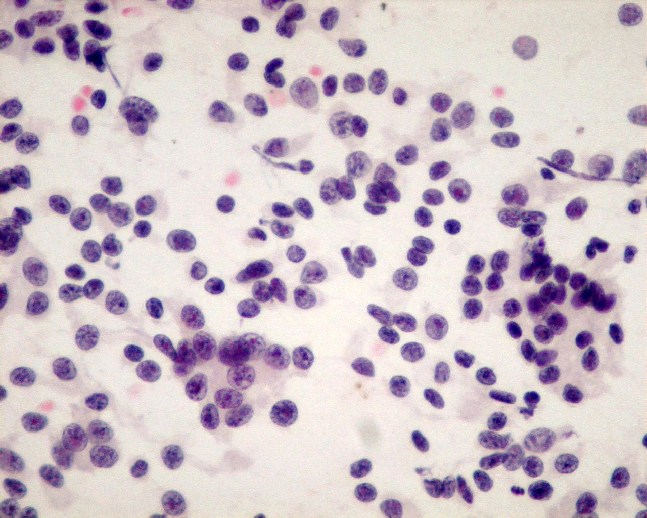
Unless you see necrosis, differential diagnosis between typical and atypical carcinoid should not be done in cytology
- Clean background
- Uniform population of round/ oval regular cells
- Single cells or loose clusters
- Acinar formations
- Finely stippled chromatin
- Small inconspicuous nucleoli
- Scant cytoplasm
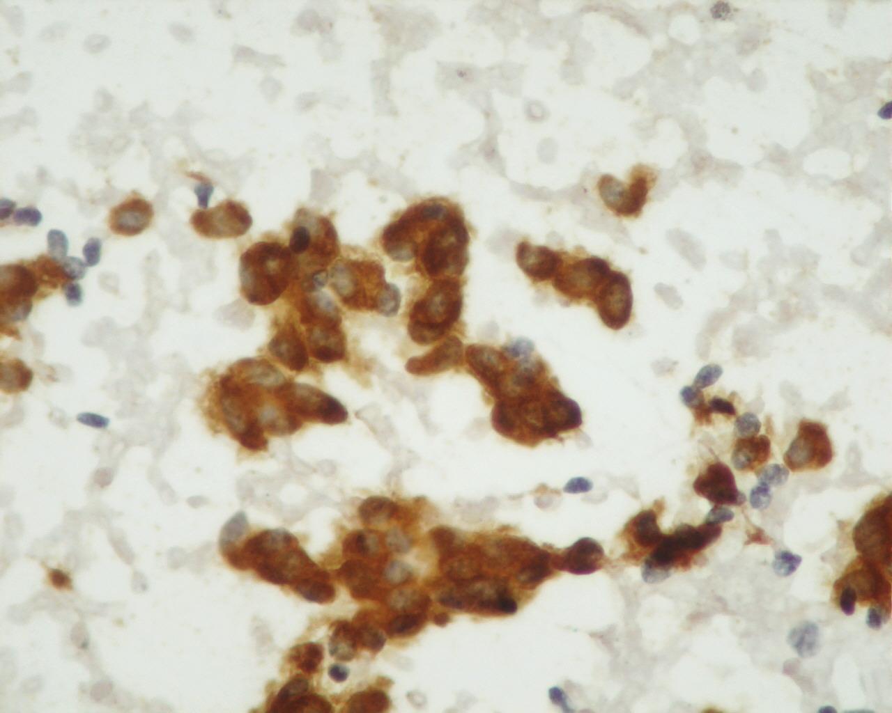
Immunocytochemistry
- AE1/AE3: Positive
- NSE: Positive
- Chromogranin A: Positive
- Synaptophysin: Positive
- CD56 (NCAM): Positive
- TTF1: Positive
Genetic studies
- Trisomy of chromosome 7
Differential Diagnosis
- Lymphoma
- Lymphoglandular bodies
- Single cells
- CD45 Positive
Main points
- Cured with resection