Clinical features
- Rare in children
- Can be congenital
- Lower extremities, buttocks, head and neck
- Deep seated and painless
- Considered as an indolent, low-grade soft tissue sarcoma with a tendency toward recurrence
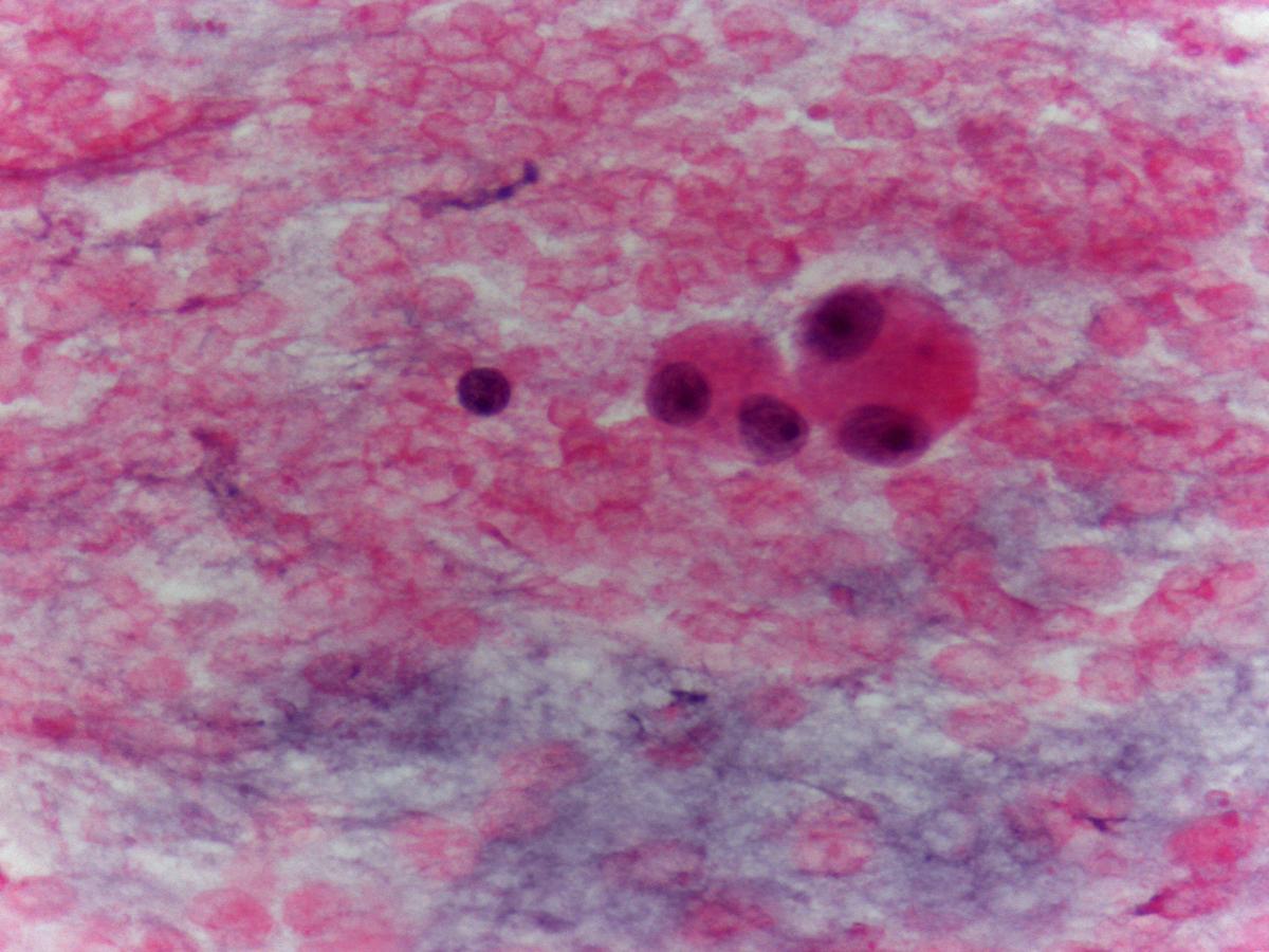
Fig 26 – Extraskeletal myxoid chondrosarcoma – Single cells or cells in small isolated groups or matrix embedded aggregates. Long delicate spicules of this matrix, with sharply defined edges, blend with the myxoid background (H&E).

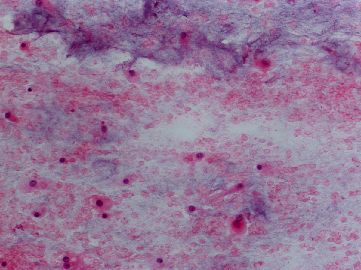
- Aspirate with abundant whitish fluid
- Metachromatic myxoid background
- Poor to rich smears
- Bimodal cell pattern: round to oval cells and spindle cells
- Single cells , cords or clusters
- Nuclei with fine granular chromatin
- Nuclear grooves can be present
- Small prominent nucleoli
- Delicate and scant to moderate amounts of pale blue cytoplasm.
- Myxoid/chondroid matrix (almost always present) embedding cell aggregates
- Fibrillary to dense stroma with sharp borders although some fragments of matrix have long delicate spicules that blend with the background
- Multinucleated cells
Histochemical Stains
- sulphated glycosaminoglycan’s stain, without staining to hyaluronic acid
- Alcian Blue PH 1
Immunocytochemistry
- S100 Protein: Positive
- Vimentin: Positive
- Lysozyme: Positive
- Bcl-2: positive
- CD99: positive
- CD57: positive
- EMA and cytokeratin’s (AE1AE3, CAM5.2, CEA): Negative
- Neuroendocrine markers: Positive
Electron microscopy
- Rudimentary cell junctions
- Cytoplasm with: well developed endoplasmic reticulum, Golgi and many mitochondria.
- RER distended and filled with parallel microtubules of 24-nm
- Cytoplasmic Glycogen
Genetic studies
- t (9; 22) (q22-31; 11-12) – EWS/TEC gene fusion-(75%)
- t(9;17)- described in some variants
Differential Diagnosis
- Rhabdomyosarcoma
- Muscle differentiation (immunohistochemical and electron microscopy)
- Myxoid liposarcoma
- Lipoblasts with scalloped nuclei clustered around branching capillaries
- Chicken wire
- Chordoma
- Also has metachromatic myxoid stroma
- Also has tumour cells single or arranged in cords
- Presence of physaliphorous cells
- Epithelial markers : Positive
- Extra renal rhabdoid tumour
- Rarely a myxoid lesion
- Prominent eosinophilic nucleoli
- Eosinophilic para nuclear cytoplasmic inclusions.
- Cytokeratin’s: Positive
Main points
- Lung metastasis are frequent at presentation, in high grade tumours