Clinical features
- Accounts for less than 10% of non-rhabdomyosarcomas in children
- Adolescents and young adults
- Male predisposition
- Painless dermal or subcutaneous nodules
- Extremities (hands, fingers, wrist and legs or genital)
- Superficial lesions
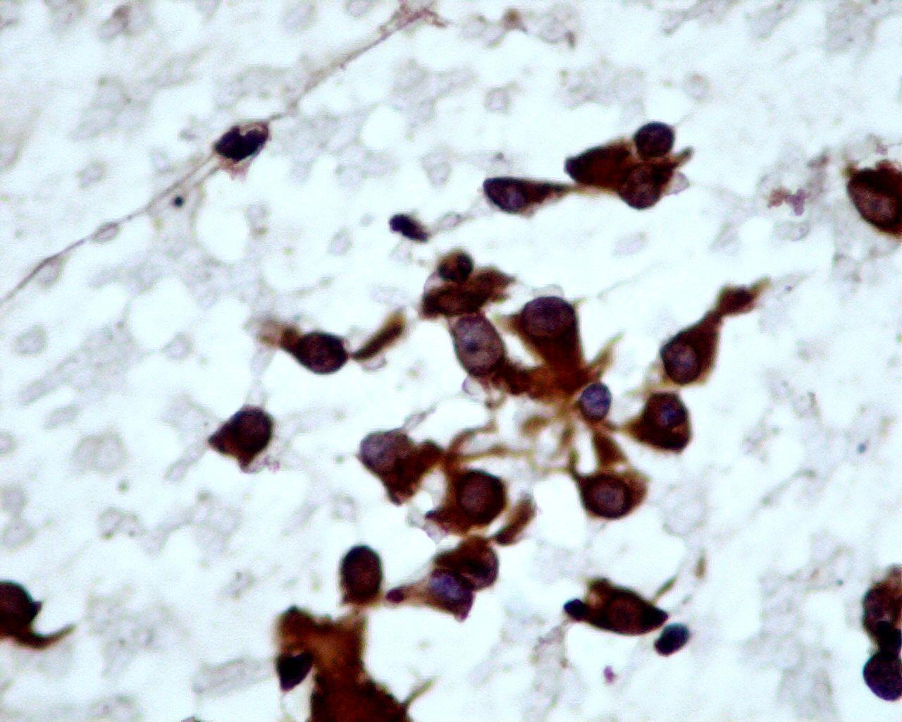
Fig 37 – Epithelioid Sarcoma – Aggregates and dispersed cells (Giemsa)
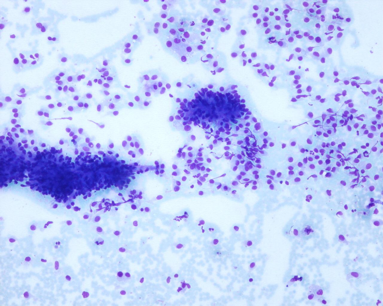
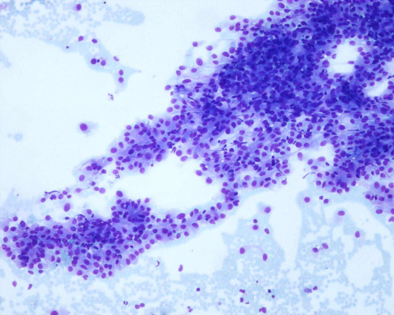
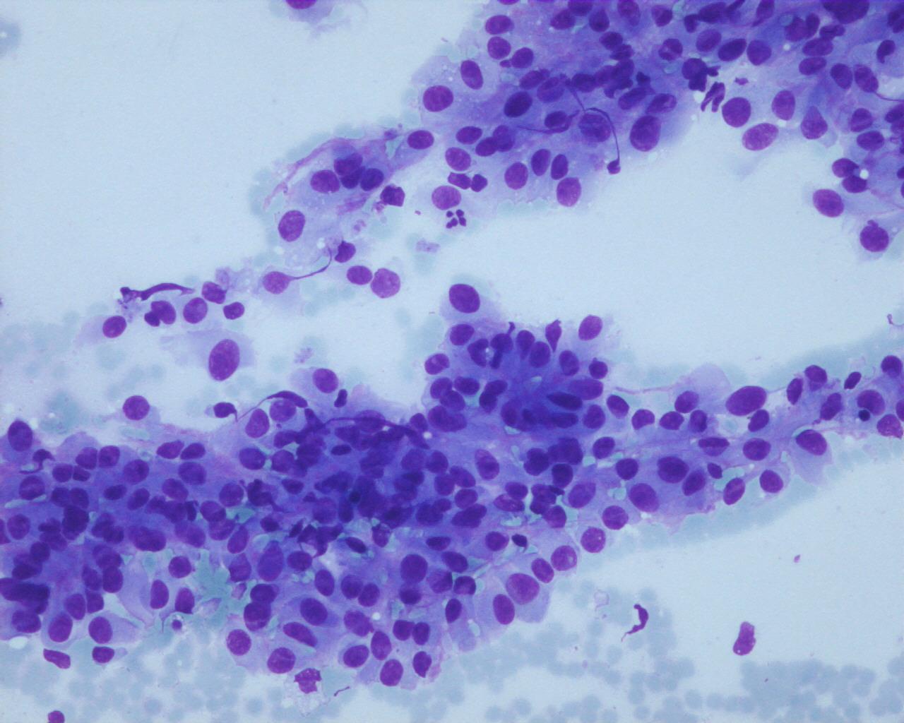
- Bloody aspirates
- Poor cellular smears
- Necrotic background
- Dispersed cell pattern or nests
- Cells with well-defined cell borders, with intercellular spaces
- Large polygonal epithelioid cells , spindle rhabdoid or multinucleated
- Irregular nuclei contour (round, oval, elongated or pleomorphic)
- Prominent nucleoli
- Dense cytoplasm with well-defined borders
- Rhabdoid inclusions
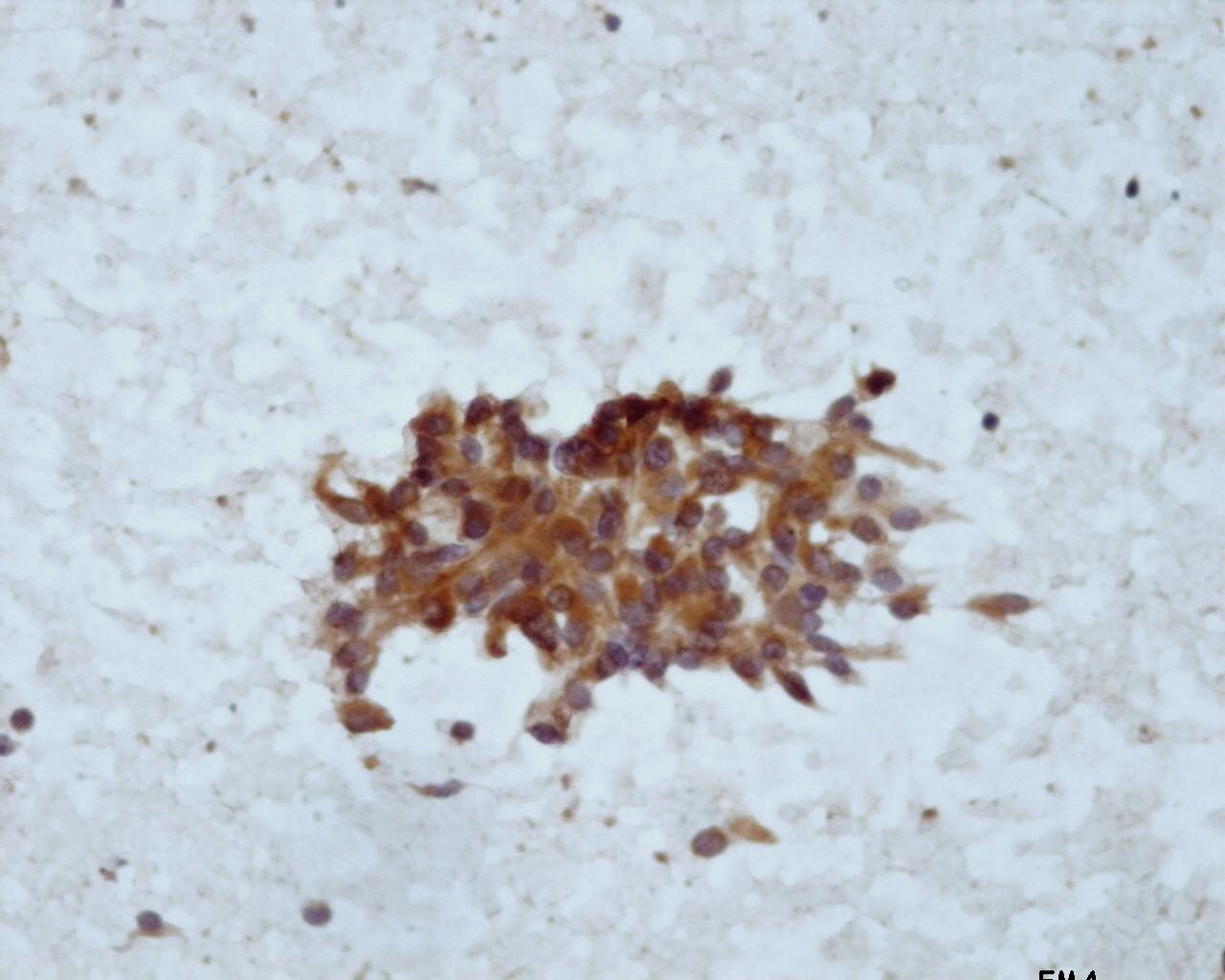
Immunocytochemistry
- Cytokeratins: Positive
- EMA: Positive
- CD34: Positive (50%)
- Vimentin: Positive (perinuclear)
- Desmin : positive
- VE-cadherin: Positive
- INI 1: Positive
- E-cadherin: Negative
- CD31: Negative (generally, rare positive cases are reported)
Electron microscopy:
- Microvilli
- Typical irregular nuclei contour and prominent nucleoli
- Cytoplasm with:
- Intermediate filaments
- Abundant polyribosome, rough endoplasmic reticulum
- Occasional lipid droplets and degenerative vacuoles surrounded by filopodia or microvilli
- Desmosome-like junctions
- Small intercellular spaces
Genetic studies:
- DNA copy number changes, gains>losses, including +11q13, 1q21-q23, 6p21.3, 9q31-qter, losses at 9pter-p23, 13q22-q32 (4)
Differential Diagnosis
- Metastatic carcinoma (squamous)
- Cells with are in syncytial clusters without the typical well-defined cell borders, and intercellular spaces of epithelioid sarcoma
- Melanoma
- S-100 protein: Positive
- HMB45: Positive
- Synovial sarcoma
- Distinction can be difficult
- CD34:Negative
- t(X:18)
- Hemangioendothelioma
- Cd31 : Positive
Main points
- Local recurrences
- Metastasis in 45% of the cases (lymph nodes ,lungs, skin )
- Poor prognostic features:
- Axial tumour
- Deep tumour
- Large size
- Haemorrhage
- Necrosis
- Mitosis
- Rhabdoid features
- Angiolymphatic invasion