Mature teratoma is not a cytological diagnosis. The presence of an immature or malignant component in a tumour cannot be ruled out through FNA.
Clinical features
- Mature tumours in children are mostly cystic (dermoid cyst)
- Bilateral in 15%
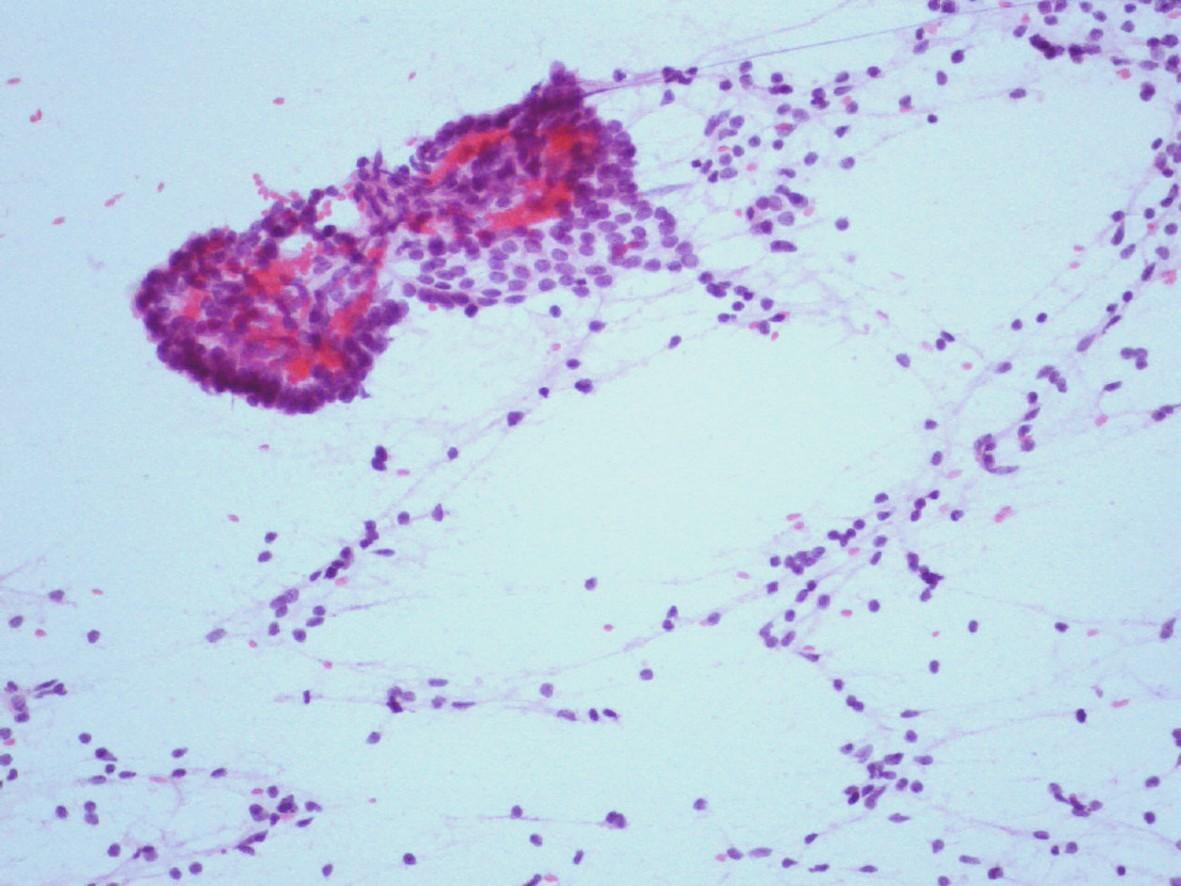
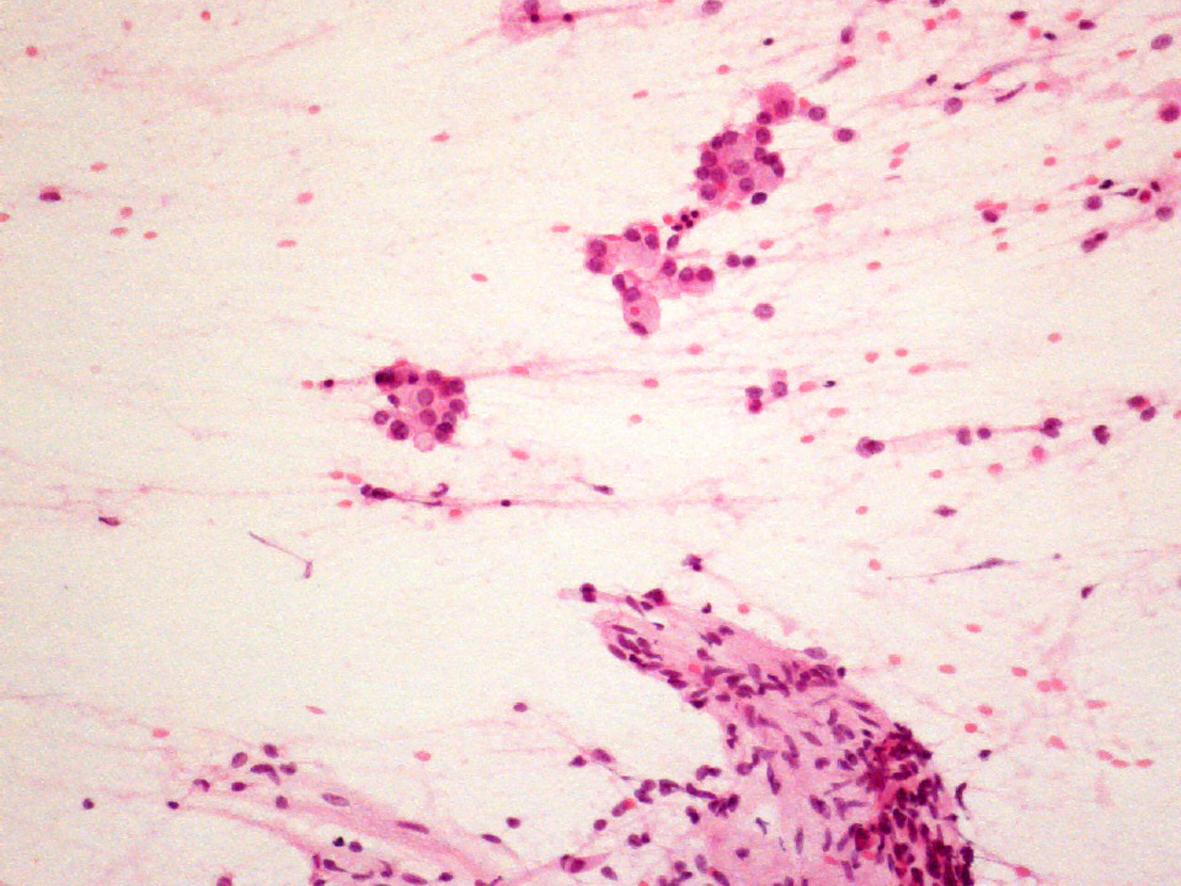
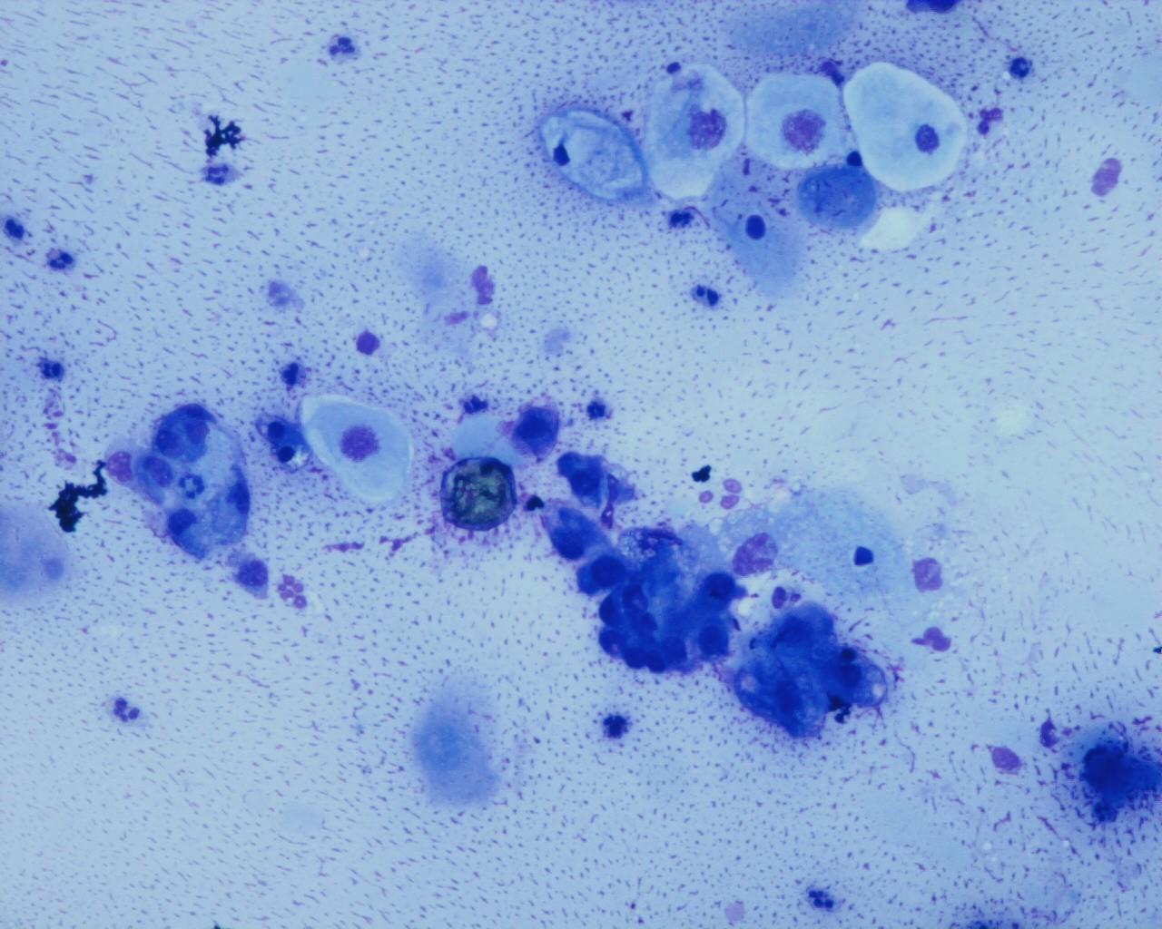
Fig 63 – Teratoma –Testicular teratoma – Adipose cells, epithelial cylindrical cells and a three-dimensional group of poorly differentiated epithelial cells. Macrophages can be seen in the background (H&E)
- Representation of the different constituents (epithelial or mesenchymatous)
- Presence of immature tissues (blastemal, cartilage, etc.)
- Presence of malignant cells
- Cystic component
Immunocytochemistry
- Stains correspond to the tissues present
Modern diagnostic techniques
- Non-contributory
Differential diagnosis
- With all the tissues and tumours that may be present in mature, immature and/or malignant teratoma
Main points
- Tumours that arise from a germ cell after first meiotic division