Clinical features
- Unusual in children (5% of the neurogenic tumours)
- Most sporadic- V and VIII cranial nerves
- Bilateral VIII cranial nerve- Neurofibromatosis type 2
- Head , neck, extremities (flexor zones)
- Slow growing and painless
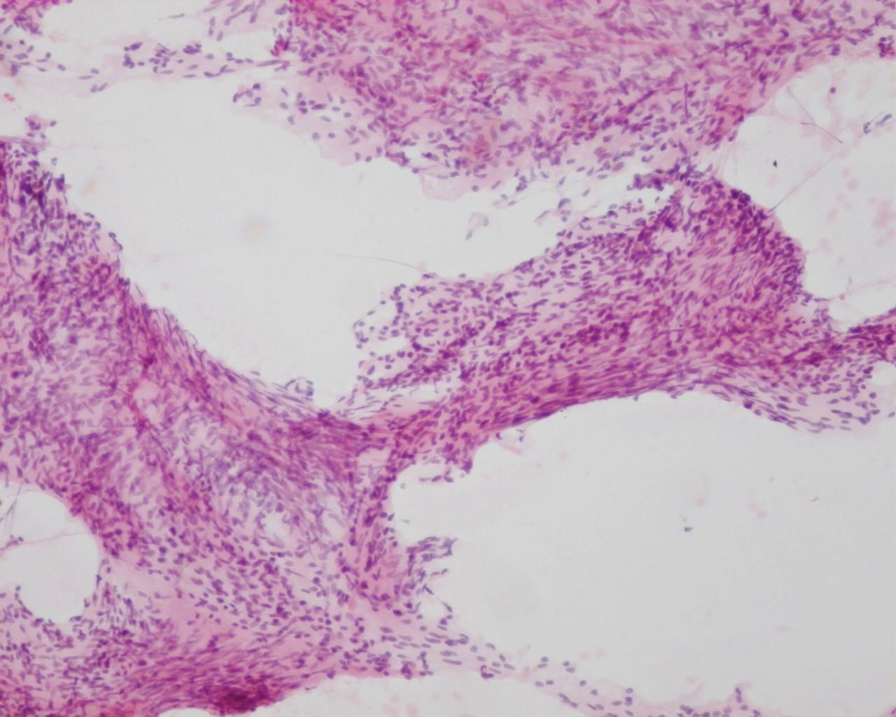
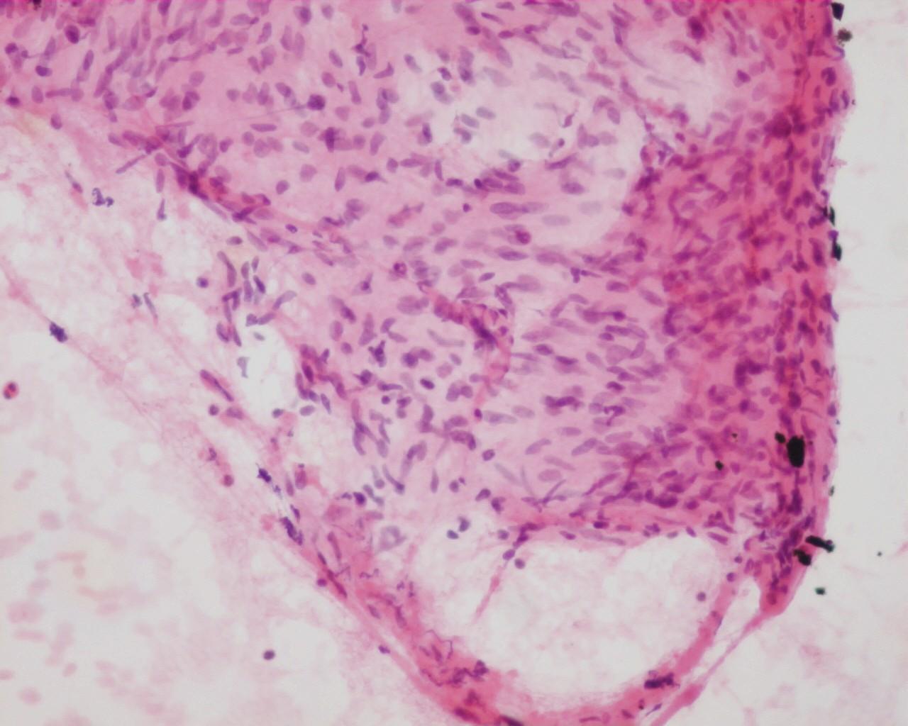
Fig 34 – Schwannoma –Cohesive tissue fragments. Fragments of uniform spindle shaped cells in a moderate collagenous matrix (H&E)
- Low sensitivity
- Pain during aspiration
- Cohesive cells in tissue fragments (helpful criteria of low grade/benign nerve sheath tumours)
- Nuclear palisading (Verocay bodies)- (helpful pattern)
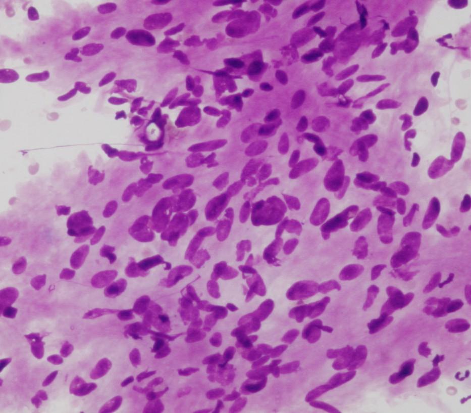
- Spindle cells with ovoid, comma or wavy nuclei
- Intranuclear cytoplasmic inclusions
- Fibrillary cytoplasm
- Anisokariosis can be present-Ancient Schwannoma
- No mitosis
- Melanotic pigment can be present in HMB45 positive cells
Immunocytochemistry
- Vimentin: Positive
- S100 protein: Positive
- Leu7: Positive
- GFAP: Positive (occasional)
Differential Diagnosis
- Neurofibroma
- Nuclear features are in most of the cases indistinguishable
- In these kind of lesions the main point of cytological diagnosis is that of a benign neurogenic tumour