logo efcs bianco

Evaluace vzorku

This content is also available in: English Español Magyar Polski Türkçe

Bookmark (0)

No account yet? Register

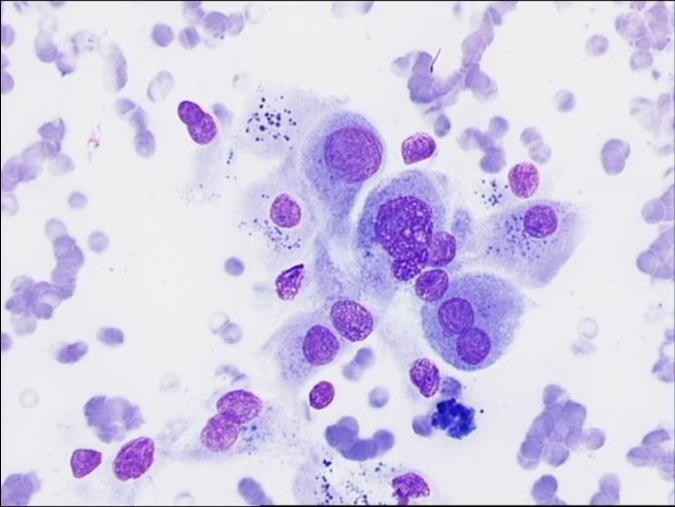
V hodnocení je t?eba popsat n?kolik znak?

typ bun?k (jako nap?.thyrocyty, makrofágy, lymfocyty)

Thyrocytes and macrophages. Fluid colloid in the background, bland thyrocytes and pigmented macrophages.
Lymphocytes and thyrocytes. A group of bland thyroid cells showing a follicular arrangement, surrounded by smaller lymphoid cells with little amount of identifiable cytoplasm. Such lymphoid cells are also inside the follicular group, suggesting chronic inflammation.

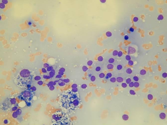
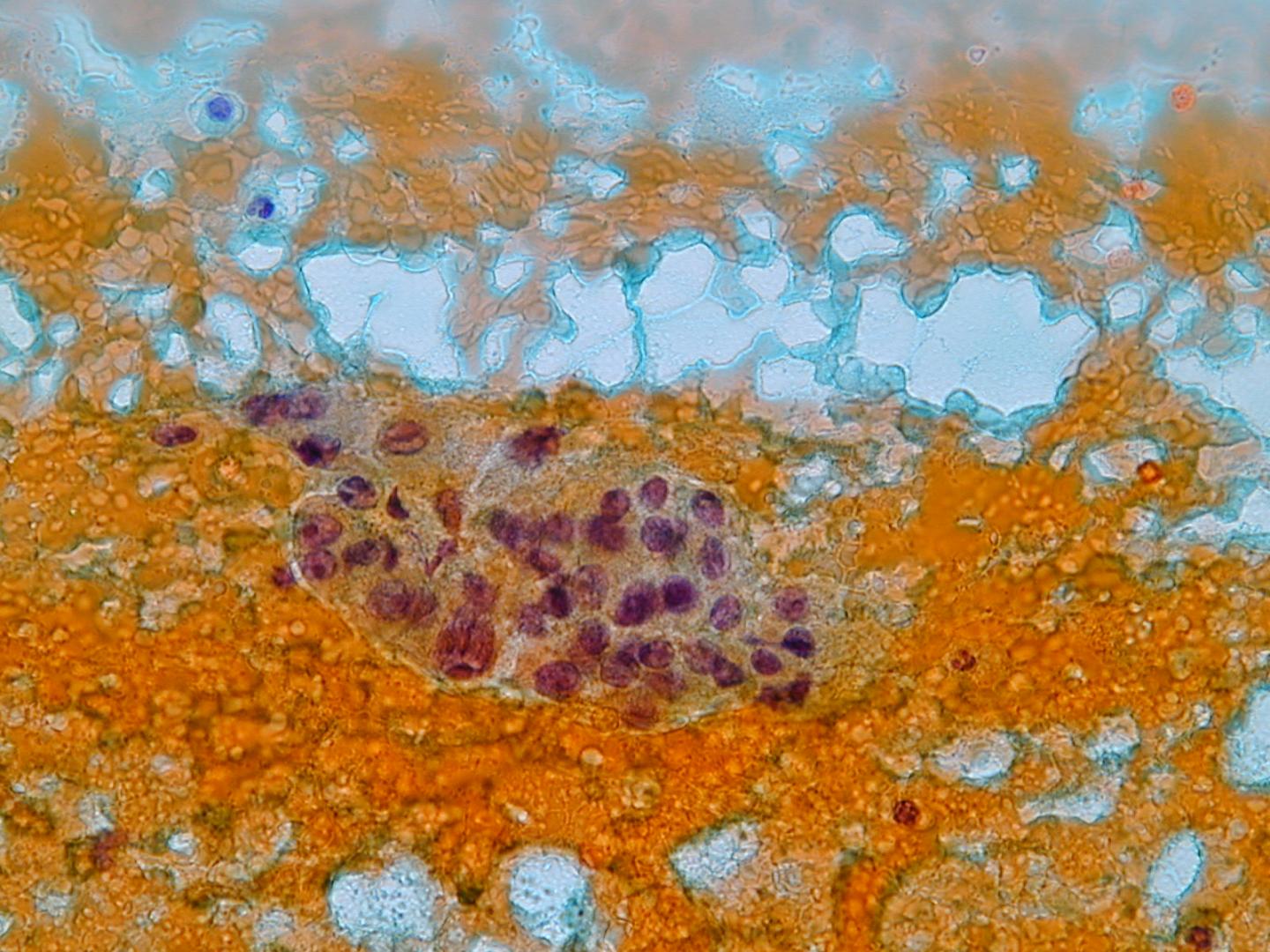
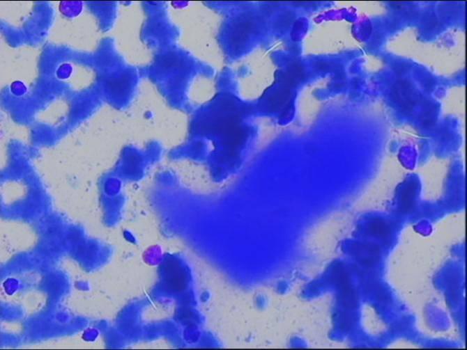
množství a kvalitu koloidu  (chudý-hojný, ?ídký-denzní)

Colloid: Fluid but relatively thick colloid with the typical cracks which are produced with air drying. These are artefacts which are useful in recognising colloid.
Colloid: A clump of thicker colloid surrounded by few bare nuclei. In the appropriate context this type of colloid may be associated with papillary carcinoma.

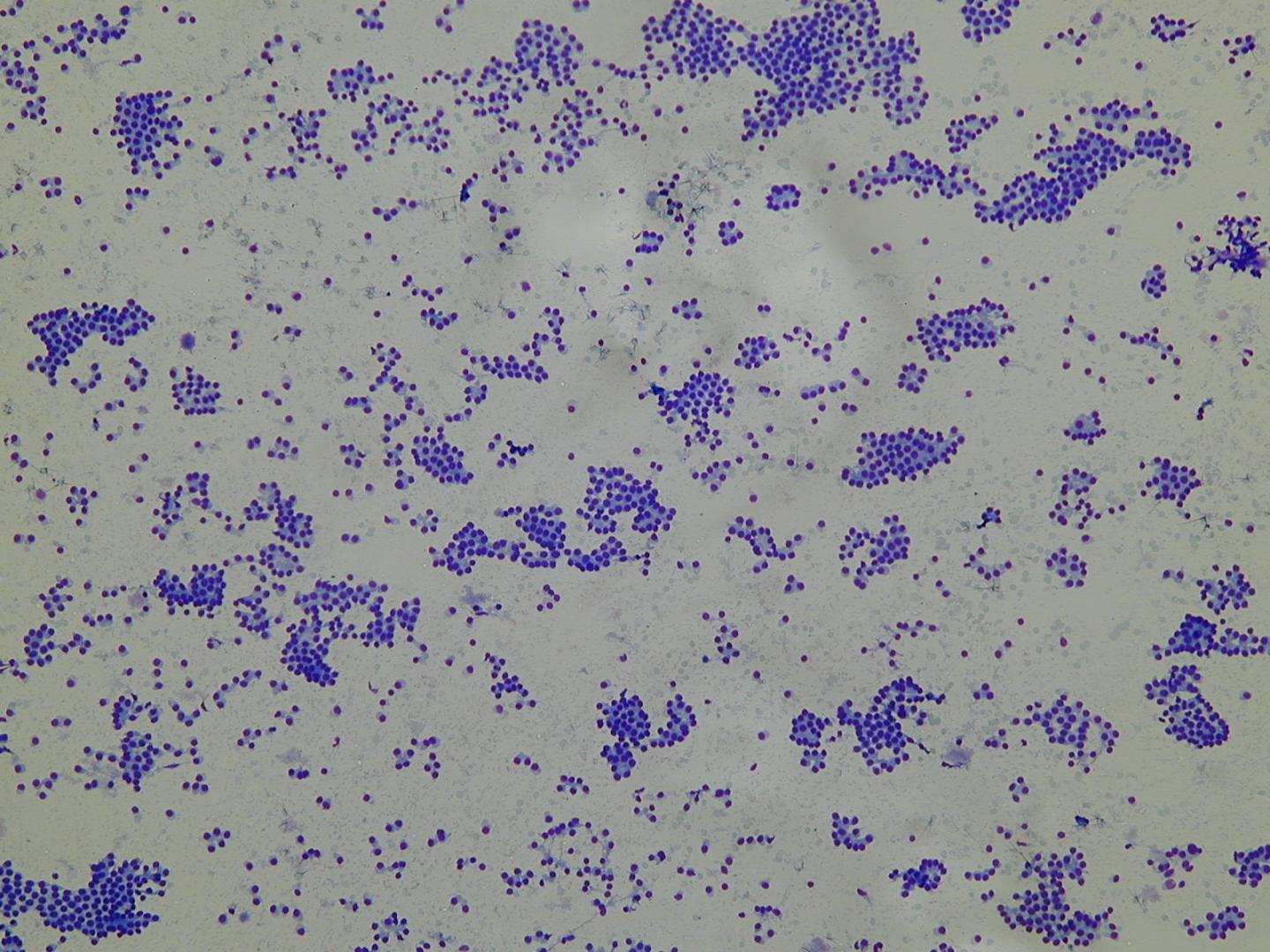
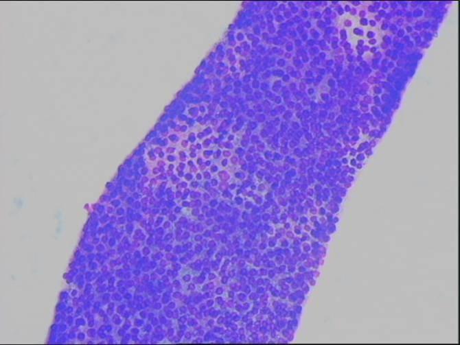
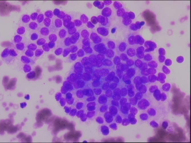
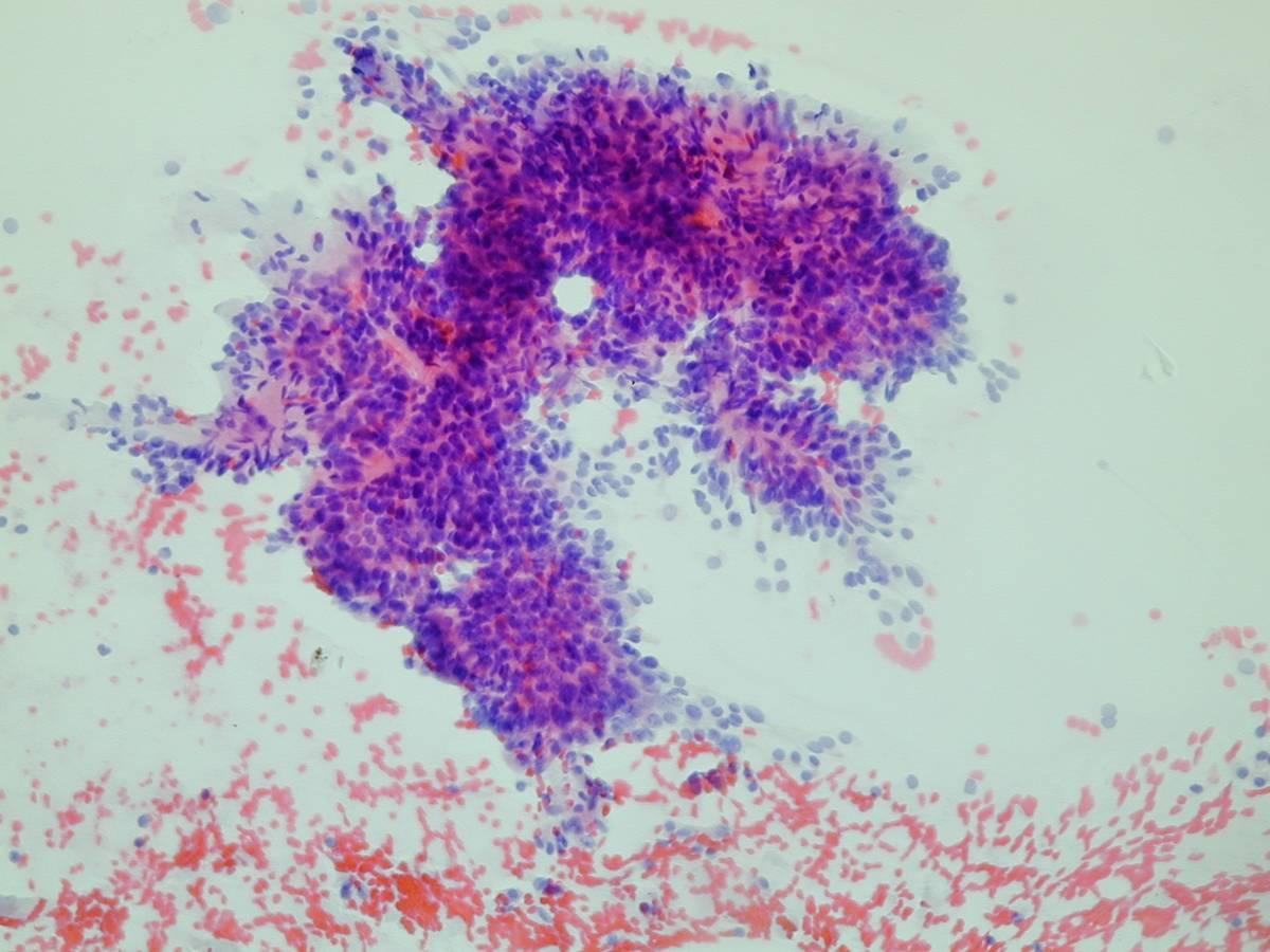
celularita (nízká, st?ední, vysoká)

Highly cellular smear showing a microbiopsy. A microbiopsy, as may be  obtained with larger needles, in which 3D follicles are separated by collagenous septa; the margins of the follicles are regular. This pattern is usually seen in FNAs from nodular goitres. Please note that in air dried samples stained with MGG the centre of these microbiopsies will be too thick to assess.
Less cellular but diagnostic smear showing groups of bland thyroid cells in a microfollicular arrangement, bare nuclei of thyroid cells and fluid colloid in the background. A few macrophages are also present. This pattern is associated with benign disease.

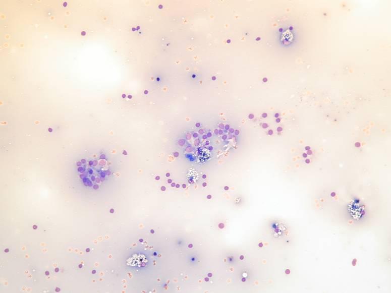
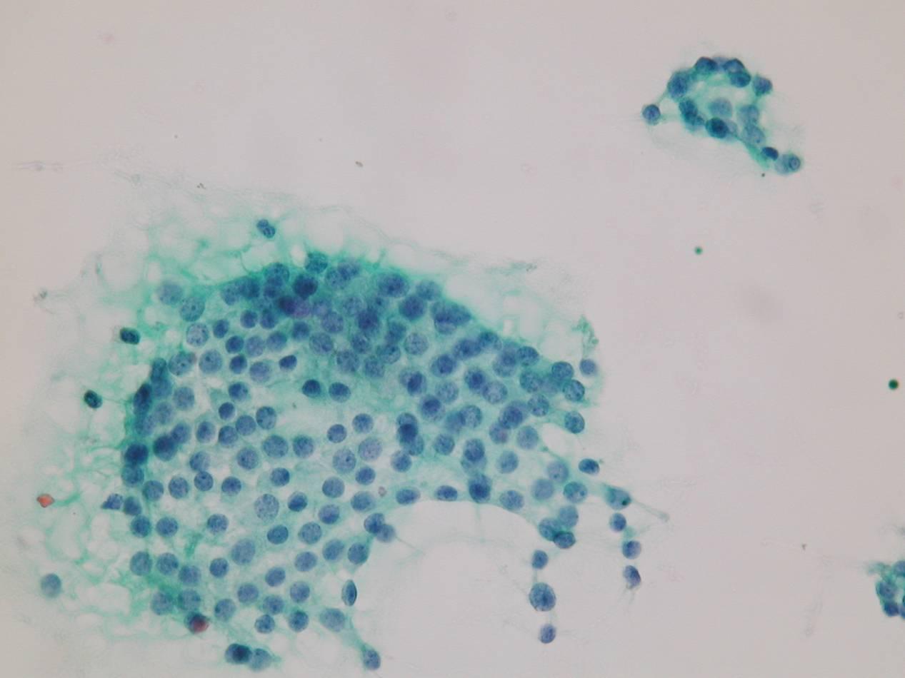
architektura (jednovrstevné formace, nat?snané trsy, plachty, makro/mikrofolikuly, papilární útržky, izolované bu?ky)

Sheets of bland thyroid cells. A monolayered sheet of bland thyroid cells. Note the regular distribution of the nuclei and cytoplasm and very fine chromatin with small nucleoli.
Sheets of bland thyroid cells. Another example of a a monolayered sheet of bland thyroid cells. Because of the size of this sheet some areas may appear more crowded but are actually simply folded. It is good practice to focus on these areas to check the regular distribution of cells along the same plane. Such large sheets are usually lining larger follicles or cysts in  benign disease.
Thyroid cells in a microfollicular arrangement. Several groups of thyroid cells in a microfollicular arrangement. In follicular lesions this may be the only type of cell present.
A papillary cluster with the epithelial cells lying perpendicular to the stromal axis. The typical nuclear features of papillary carcinoma need to be present however for a diagnosis of papillary carcinoma..

p?ítomnost holých jader

Many bare nuclei are seen dispersed together with bland thyroid cells in a follicular arrangement. Note that the chromatin pattern is the same, confirming that these nuclei have been stripped of their fragile cytoplasm during the smearing procedure. Lymphocytes are smaller, have a more granular chromatin, folded  nuclear membrane and scanty but clearly identifiable, dense cytoplasm.

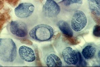
Cytologické rysy (cytoplazma, jádra)

V nádorových uzlech jsou vzorky zpravidla vysoce bun??né. Plošné formace jsou b?žné v hyperplastické strum? a makrofolikulárních adenomech, mohou však být p?ítomny i v karcinomech. Makrofolikuly jsou obvykle sou?ástí polynodózní strumy a makrofolikulárních adenom?. P?evaha  mikrofolikul? indikuje folikulární neoplázii. Papilární trsy s bu?kami na povrchu fibrovaskulární složky jsou charakteristické pro papilární karcinom. Nát?ry s p?evahou koloidu oproti folikulárním bu?kám indikují benigní uzel.

Cytologické rysy:

  • cytoplazma (množství, barvitelnost)
  • struktura chromatinu
  • jaderná membrána (hladká nebo nepravidelná)
  • jaderné rýhy a pseudoinkluze
Abundant cytoplasm containing granules. A group of large non cohesive cells with abundant dense, cytoplasm containing granules. The nuclei are single or multiple and pleomorphic. The differential includes Hürthle cells (oncocytic change), “squamoid” cells in papillary carcinoma or goitre and atypical cells in anaplastic carcinoma.
Nuclear pseudoinclusions and groovings. A cluster of large cells with abundant, polygonal and dense cytoplasm; the nuclei in this case show all the features of papillary carcinoma (nuclear pseudoinclusions, grooving).