logo efcs bianco

Mucoepidermoid carcinoma

This content is also available in: Español Português Čeština Magyar Polski

Bookmark (0)

No account yet? Register

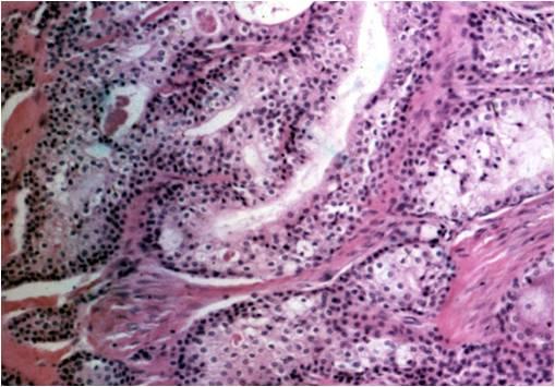
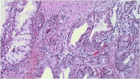
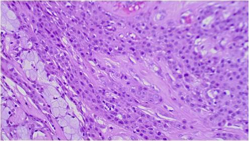
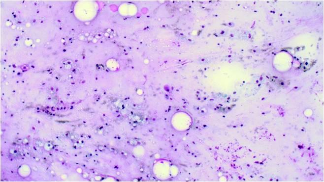
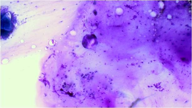
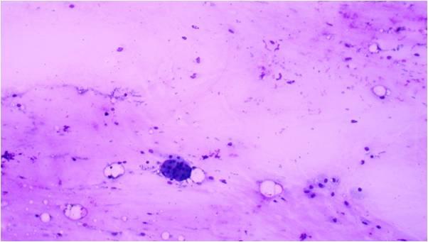
This tumor consists of squamous, mucus-producing and intermediate cells embedded in a mucinous background. It is the most common primary salivary gland malignant tumor in both children and young adults. Low, intermediate and high grade tumors are distinguished in histology on the basis of the presence of a cystic component, necrosis, neural invasion, proportion of epidermoid cells as compared to mucinous ones, anaplasia and mitotic count.

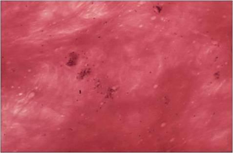
The cytological smear is sometimes very typical: all the cell types are found embedded in a mucinous background. In some cases atypia are mild, oncocyte-like epidermoid cells predominate in a dirty, ‘thick’ background, resembling a Whartin tumor. Since the mucoid material get easily into the puncturing needle even without containing epithelial cells, it is one of the most difficult cytological diagnoses in the salivary glands!

The histology is simple, usually even very typical for mucoepidermopid cc.
One of the most difficult diagnosis in salivary gland cytopathology.The age is not typical, the tumor is in almost every case multifocaL The latter is resulting in quite a few normal epithelial cells as well. The mucinous content may be abundant ( see picture below, stained with H&E ), the epithelial elements only slightly atypical. There are many other lesions showing the presence of the same „mixture”: mucus, squamous and secretory glandular cells mixed up with normal salivary gland cells ( inflamed Warthin’s tumor, metaplastic inflammatory changes, etc. ). In the image on the right lower corner a huge amount of epithelial mucus is seen with small groups of epithelial elements. The image as such does not suggest malignancy at all! In dedifferentiated cases with squamous dominance the „false” diagnosis is usual squamous cc. (H&E stain)


In the histology the epidermal component is highly atypical and dominating (H&E stain)
 
The FNAC smear shows scant cellularity, mostly euchromazic mucous background and as sign of infiltration even acinic cells. (Giemsa stain)