logo efcs bianco

Autoimmune sialoadenitis (Benign lymphoepithelial lesion)

This content is also available in: Español Português Čeština Magyar Polski

Bookmark (0)

No account yet? Register

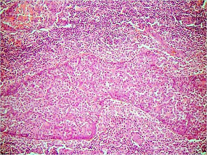
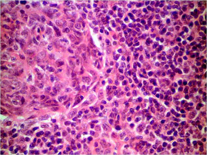
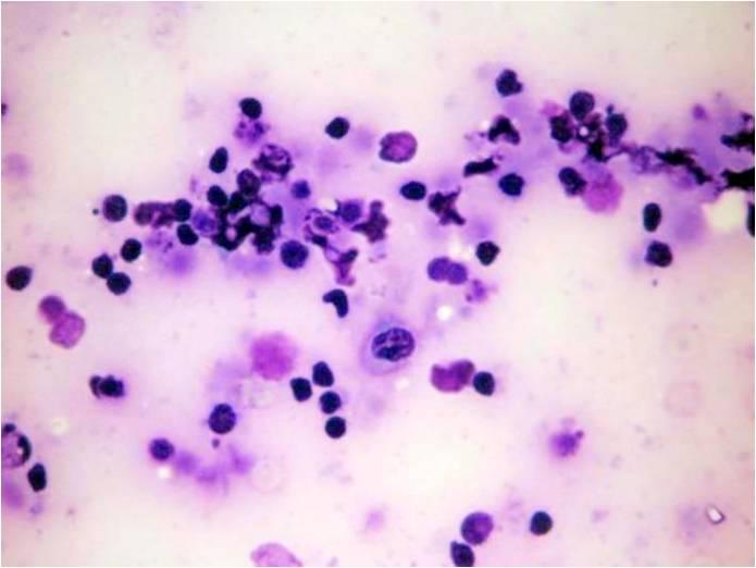
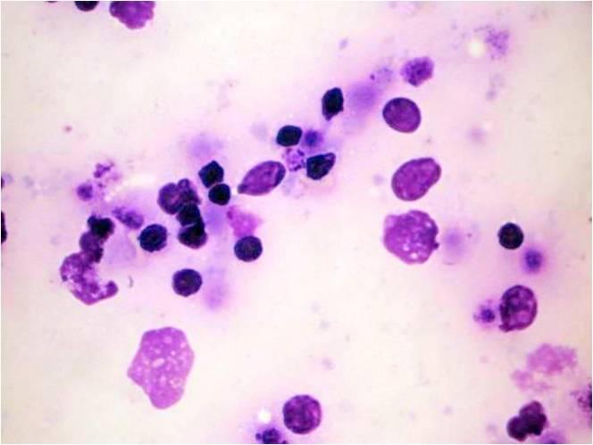
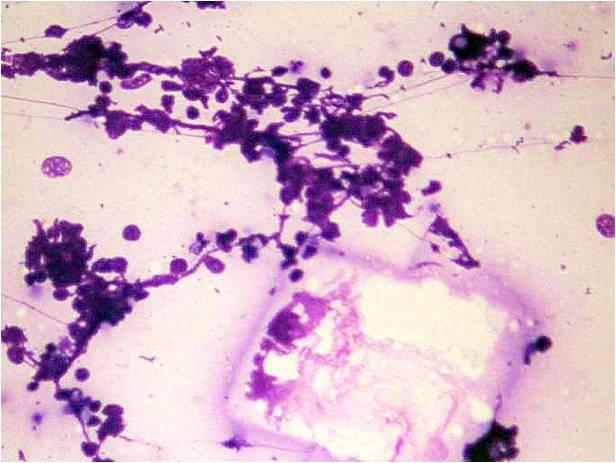
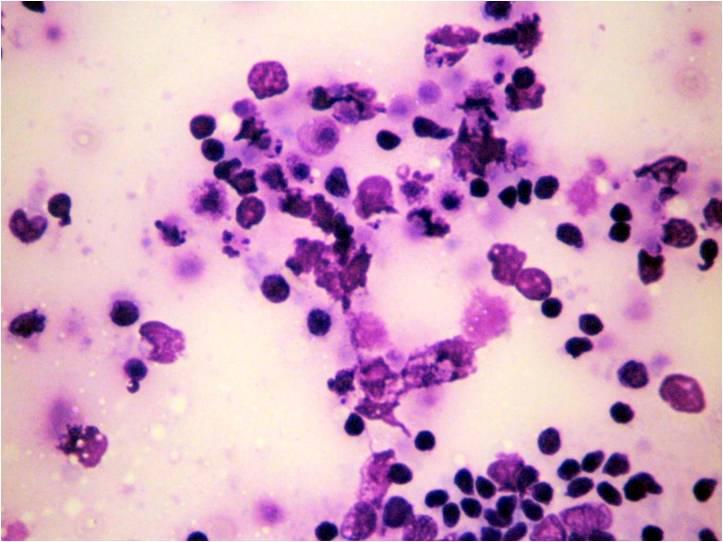
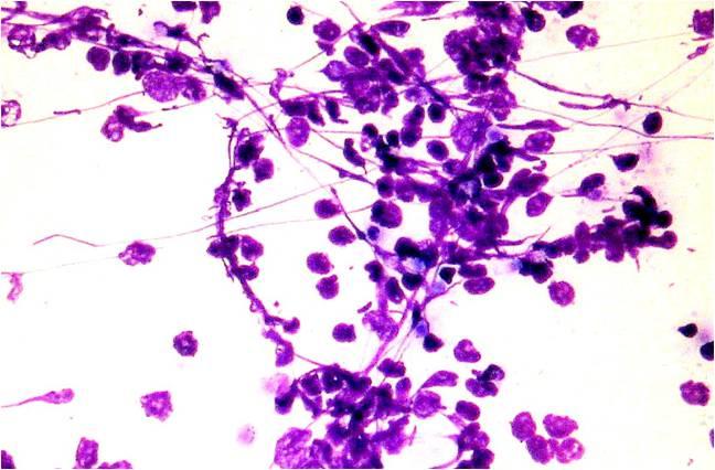
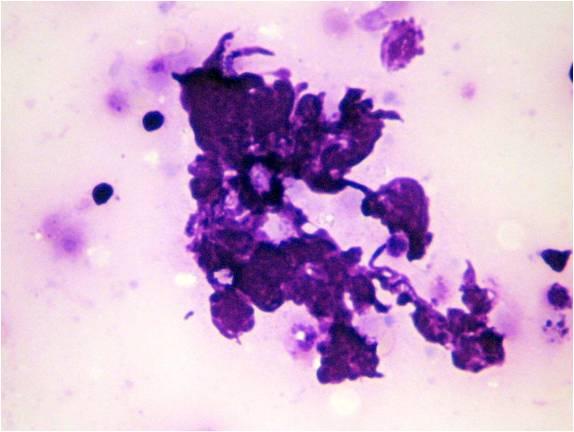
The smear looks like the one from a lymph node: small lymphocytes, follicular lymphocytic cells, epithelioid cells, plasma cells dominate. Salivary gland originated epithelial cells are also present, embedded in the above background. The lesion is often cystic; in this case the aspirate is fluid. Acinic cells of the surrounding area and proliferative ductal cells are present. The epithelial cells may show osmotic changes: ill-defined cytoplasm, larger nucleus which is pale stained. In case of follicular lymphoid proliferation the differentiation from malignant lymphoma may be difficult. The lesion may be connected or part of   the Sjögren Syndrome.

Histology: Destructive lymphoid tissue occupying the salivary gland with epi-myoepithelial islands  


Cytology fo the autoimmune sialoadenitis: destruction is represented by cystic change; lymphoid tissue is dominating the picture ; cristalloid as part of the cystic content is also visible 


Cytology of the autoimmune sialoadenitis: destruction is represented by cystic change; lymphoid tissue is dominating the picture ; the cells are highly vulnerable