logo efcs bianco

Adenoid cystic carcinoma

This content is also available in: Español Português Čeština Magyar Polski

Bookmark (0)

No account yet? Register

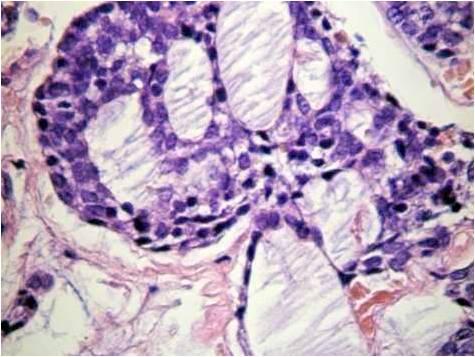
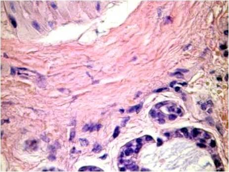
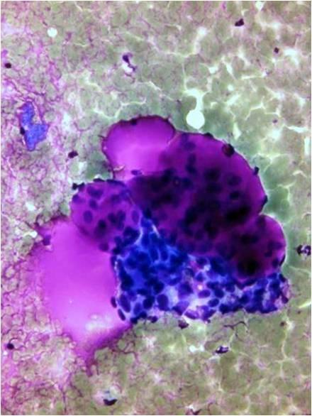
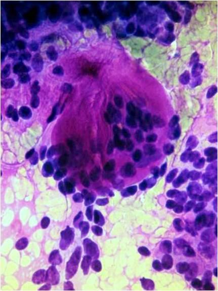
This is an infiltrative ‘basaloid’ tumor, which is the most common malignant tumor in the salivary glands. It is a slow growing but highly malignant carcinoma. The appearance is similar to that of the cylindroma of the sweat glands in the skin; however the tumor localized to the skin is regarded as benign, whereas the same pattern is predictive of malignant behavior in the salivary glands. It is a tumor of the 5th – 6th decades. Pleomorphic adenomas may show the presence of ‘adenoid cystic areas’, but this is not predictive of malignancy in mixed tumor! Histologic pattern is cribriform, tubular or solid. The pattern is always characterized by a perineural infiltrative growth. Cytological smears are rich in basaloid, monomorphic cells (the cytomorphology does not correspond to that of a highly malignant tumor!), closely connected to or embedded into metachromatic, cylindriform hyaline globules. The basaloid cell population is most probably myoepithelial in origin.


Histology (H&E): The epithelial component of this tumor is usually basaloid, very monomorphous, not at all suggesting malignancy! The histology is a peculiar, well known lesion, often accompanied by perineural invasion. The Giemsa stain is again very much helpful in diagnosing this lesions: large cylinders are seen, which are partly surrounded partly covered by the epithelial elements. The admixture of normal salivary gland cells is usually not typically present. These tumors behave highly malignant in the salivary glands, benign in the skin. 
Cytology (Giemsa): The epithelial component of this tumor is usually basaloid, very monomorphous, not at all suggesting malignancy! The histology is a peculiar, well known lesion, often accompanied by perineural invasion. The Giemsa stain is again very much helpful in diagnosing this lesions: large cylinders are seen, which are partly surrounded partly covered by the epithelial elements. The admixture of normal salivary gland cells is usually not typically present. These tumors behave highly malignant in the salivary glands, benign in the skin. 
Cytology (Giemsa): The epithelial component of this tumor is usually basaloid, very monomorphous, not at all suggesting malignancy! The histology is a peculiar, well known lesion, often accompanied by perineural invasion. The Giemsa stain is again very much helpful in diagnosing this lesions: large cylinders are seen, which are partly surrounded partly covered by the epithelial elements. The admixture of normal salivary gland cells is usually not typically present. These tumors behave highly malignant in the salivary glands, benign in the skin.