logo efcs bianco

Warthin tumor

This content is also available in: Español Português Čeština Magyar Polski

Bookmark (0)

No account yet? Register

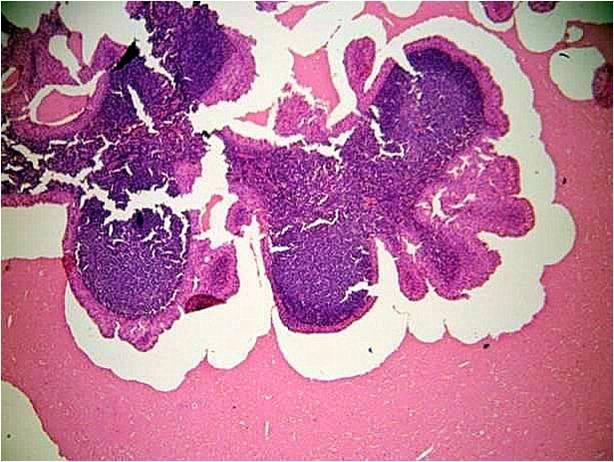
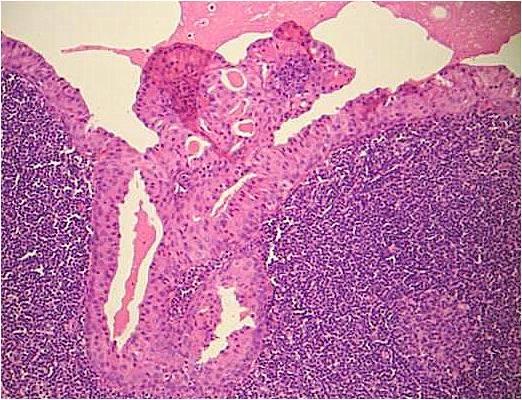
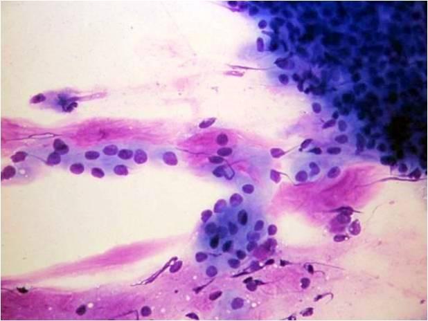
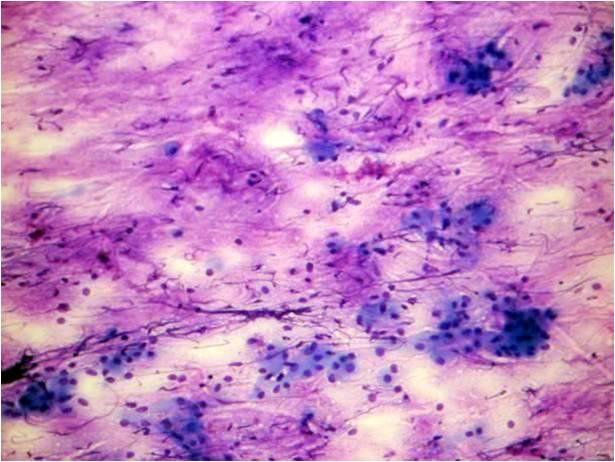
It occurs mainly in males, it is frequently bilateral. It has a cystic, oncocytic lymphoepithelial appearance. The cytology is that of a cystic lesion. The aspirated material is usually brown, dirty and thick. The background of the smear is composed by this ‘cystic’ dirty fluid, which contains oncocytic cells, lymphocytes and histiocytes. The oncocytes are in papillary or pseudopapillary structures. The Whartin tumor (cystadenoma papillare lymphomatosum) frequently undergoes necrotic changes. This results in squamous metaplasia. The latter may mimic necrotic squamous cancer. The differential diagnosis very important, since squamous carcinoma in the head and neck region is usually necrotic, hence pseudocystic. Nearly all of the patients are smokers.

Typical histology of the Whartin tumor ( cystadeno-lymphoma ): smaller/larger cyst, line by two rows of oxyphil ( oncocite like ) cells. The content is eosinophil, more or less homogeneous according to the size and the grade of inflammation. The epithelial elements are intimately connected to the lymphatic background. The lesion is often multiple, commonly seen in lymph nodes as well. It is easy to understand, that using vacuum (FNAB) the less „fixed” component is the fluid. This will be followed by the lymphocytes and the oncocytic epithelial elements.(see next views! ) (Giemsa stain
Typical histology of the Whartin tumor ( cystadeno-lymphoma ): smaller/larger cyst, line by two rows of oxyphil ( oncocite like ) cells. The content is eosinophil, more or less homogeneous according to the size and the grade of inflammation. The epithelial elements are intimately connected to the lymphatic background. The lesion is often multiple, commonly seen in lymph nodes as well. It is easy to understand, that using vacuum (FNAB) the less „fixed” component is the fluid. This will be followed by the lymphocytes and the oncocytic epithelial elements.(see next views! ) (Giemsa stain)


Typical cytology of the Whartin tumor ( cystadeno-lymphoma ): large amount of cystic fluid ( which look like purulent for being thick and yellowish at the syringe! ) oxyphil ( oncocite like ) cells in sheets. On the right side many lymphocytes are seen. The cystic content may or may not be metachromatic, but the epithelial part is nearly always monomorphic ( as compared to the mixed tumor). In case of heavy inflammation, rupture of some of the cystic lesions, squamous metaplasia may occur, which suggests the diagnosis of a mucoepidermoid carcinoma ( mucinous background, glandular and squamous epithelium!) The FNAB of this lesion if clearly showing us how shell we expect and foresee the cytology based apon the histology: the amount of the different components of the lesion is consequence of their wither stand to the vaccum! Giemsa stain)