This content is also available in:
![]() Español
Español ![]() Português
Português
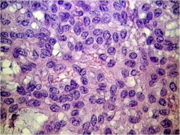
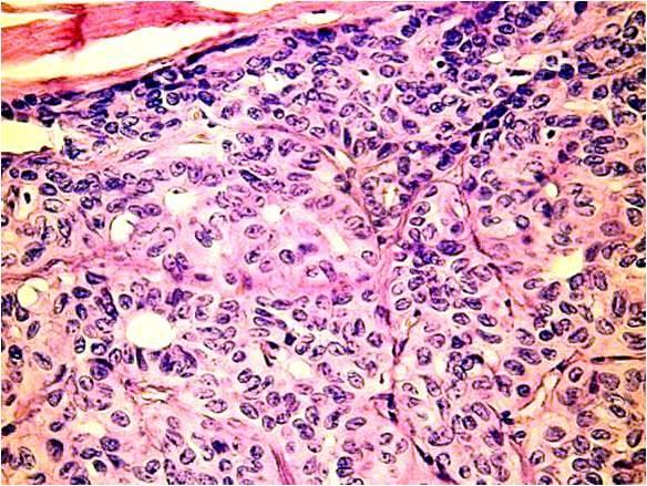
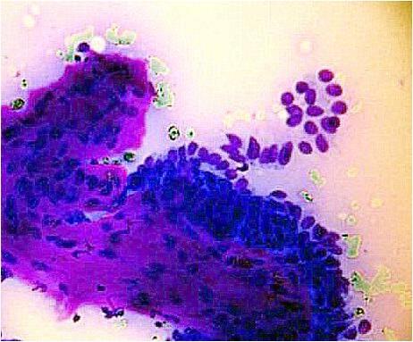
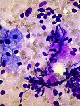
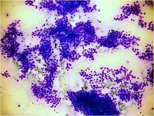
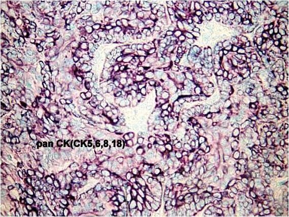
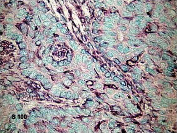
The tumor is composed of two cell types which form duct-like structures: ductal cells (inner layer), clear myoepithelial cells (outer layer). They can be distinguished by immunocytochemistry: the former are pan-keratin positive, the latter are S-100 positive. The cytology is that of a malignant clear cell tumor, with varying degrees of cellular and nuclear atypia. There are both tissue fragments and isolated cells; the cells may be arranged in solid and papillary structures. The stromal component is metachromatic, with a hyaline, elongated appearance. In some occasions duct-like or globular structures are also found. The cytological pattern suggests a malignant growth.
Epimyoepithelial carcinoma
Malignant myoepithelioma
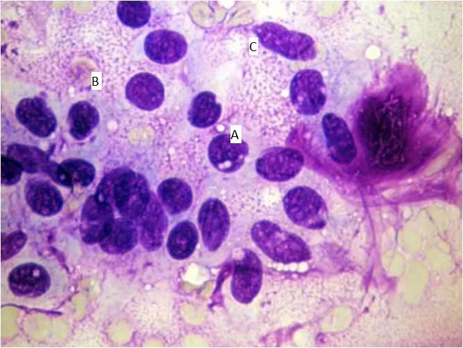
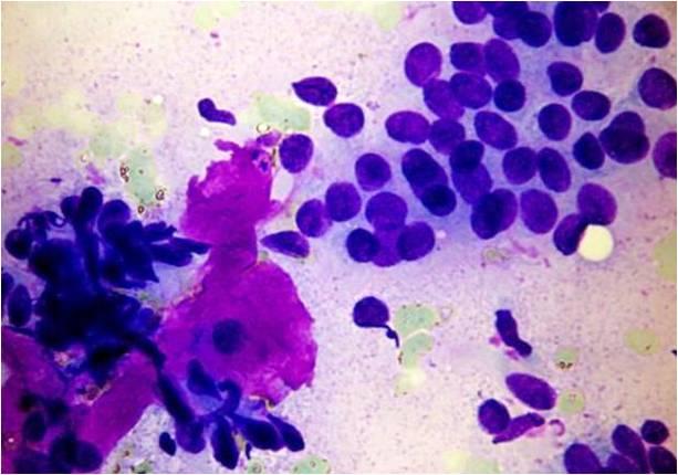
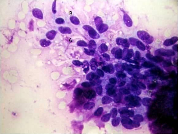
The cytology shows enlarged cells, with large ovoid nucleus, uneven chromatin. Sometimes squamoid differentiation is taking place (A) (Giemsa stain).The cells are vulnerable, as a consequence of producing the smear, elongated, irregularly shaped nuclei will be produced (B).
The cytology shows enlarged cells, with large ovoid nucleus, uneven chromatin. Sometimes squamoid differentiation is taking place (A) (Giemsa stain).The cells are vulnerable, as a consequence of producing the smear, elongated, irregularly shaped nuclei will be produced (B).