This content is also available in:
![]() Español
Español ![]() Português
Português ![]() Čeština
Čeština ![]() Magyar
Magyar ![]() Polski
Polski
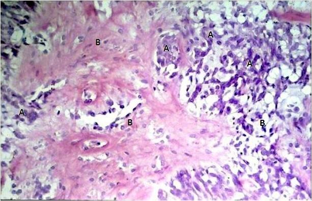
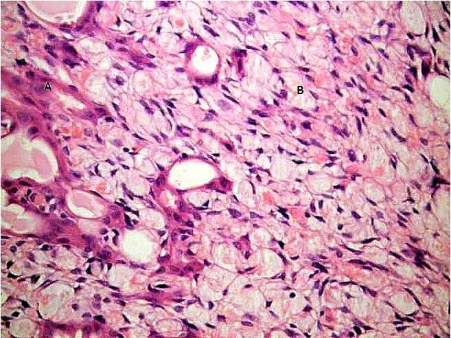
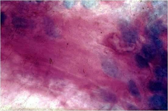
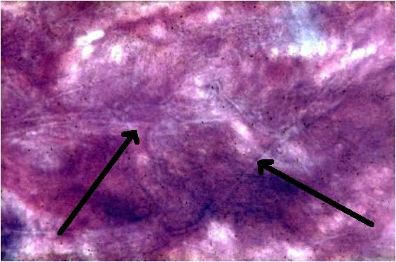
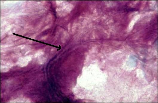
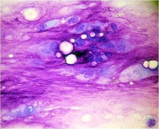
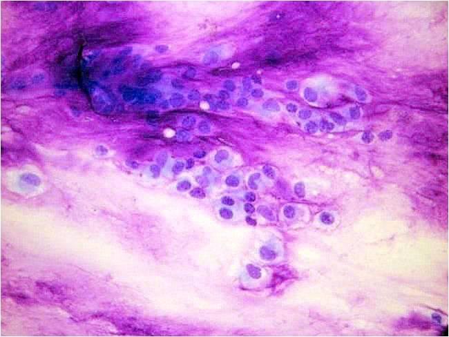
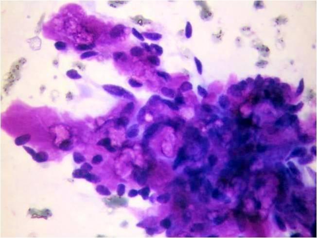
This tumor is also called pleomorphic adenoma for its composite structure. It consists of myoepithelial elements, in some cases differentiating into squamous keratinizing cells or glandular structures, together with a stromal component. The myoepithelial cells may be plasmacytoid or epithelioid. Formation of cysts may occur. The stroma may be chrondroid, hyalinized, fibrotic or even osseous, usually prettyacellular. It is the most common tumor of the salivary glands comprising 60 70% of all salivary gland tumors, and arising more often in women. The complex structure of the tumor gives rise to the complex, sometimes polymorph cytological picture. Using the Giemsa stain the chondromyxoid stromal component is metachromatic enabling a simple diagnosis. The chondromyxoid stroma shows the presence of thin, pale strands in an irregularly organized pattern. This kind of stroma is diagnostic and seems to be very similar to the stroma found in the rare tumor called chordoma, which may be located to the nasopharyngeal area. The above mentioned strands are usually not seen in adenoid cystic carcinoma. A few atypical epithelial cells do not refer to malignant transformation; still there is a known in situ malignant mixed tumor, with groups of evidently atypical epithelial cells, mixed up with myoepithel. The more monomorphic the cellular picture is the more is it suggestive of early malignant change.
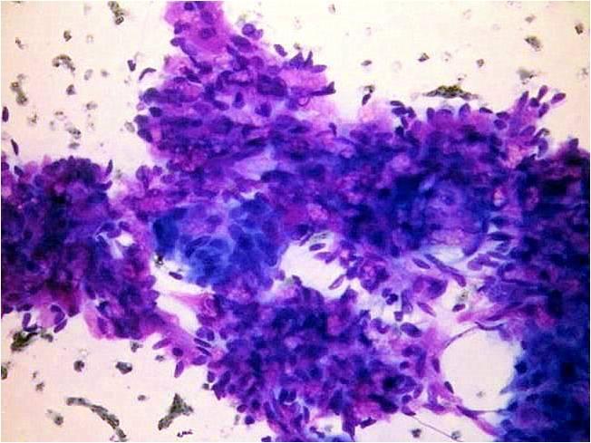
8 9% of mixed tumors may undergo malignant transformation after 15 – 25 years; the result is an invasive, highly malignant tumor, usually a squamous cell or undifferentiated carcinoma. In very rare occasions spindle cell sarcomatous tumors or even carcinosarcoma can be found. The cytological malignancy of these tumors is usually simple to recognize.