logo efcs bianco

Acinic cell carcinoma

This content is also available in: Español Português Čeština Magyar Polski

Bookmark (0)

No account yet? Register

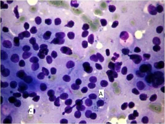
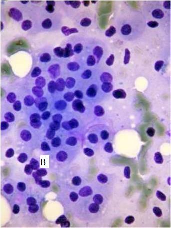
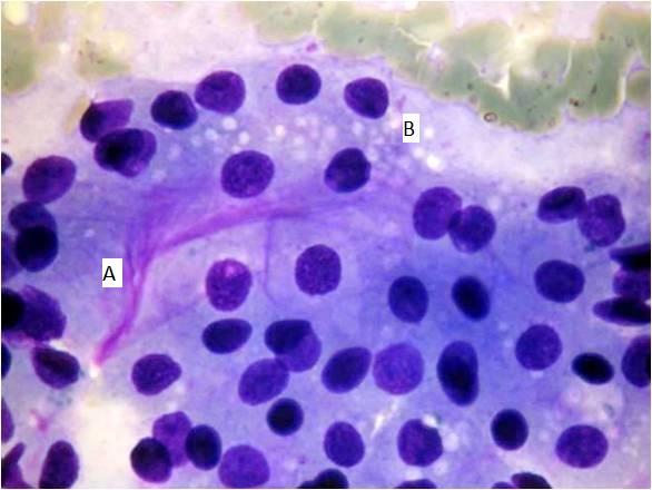
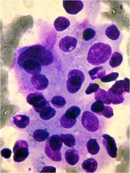
Acinic cell carcinoma occurs most frequently in the parotid gland. The former name was ‘acinic cell tumor’ because in some cases only very mild atypia is present. 2 – 7% of all salivary gland tumors belong to this group.

There are four patterns: solid, microcystic, papillary-cystic, follicular. Four cell types are usually found in this tumor: acinar cells, clear cells, intercalated duct-like cells, vacuolated cells. All patterns and all cells are usually present in the same tumor, some of them may predominate. The tumor is more common in women and it may occur in very young age as well. The different cell types are identified in the cytological smear. The tumor is usually infiltrated by many lymphocytes, which is an important diagnostic feature. In this case the cytological picture is similar to that of a well-differentiated adenocarcinoma metastatic to a lymph node. In case well-differentiated acinic cells are seen without any ductal elements, you always have to think of an acinic cell carcinoma, even if the patient is very young.

Typical acinic cells dominate the picture. The cytoplasm is granular (A), vacuolated cells (B) and intercalated cells( (C ) are also visible. There a a few lymphocytes also present, this feature is sometimes expressed in a way, that the whole image is suggesting the puncture of a metastatic lymph node. Not always all the cell types are to be found. The more cell types are present  the mores stable is the diagnosis. Ductal structures are absent!