logo efcs bianco

Wole wieloguzkowe

This content is also available in: English Español Čeština Magyar

Bookmark (0)

No account yet? Register

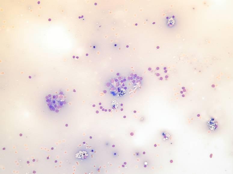
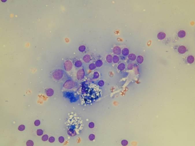
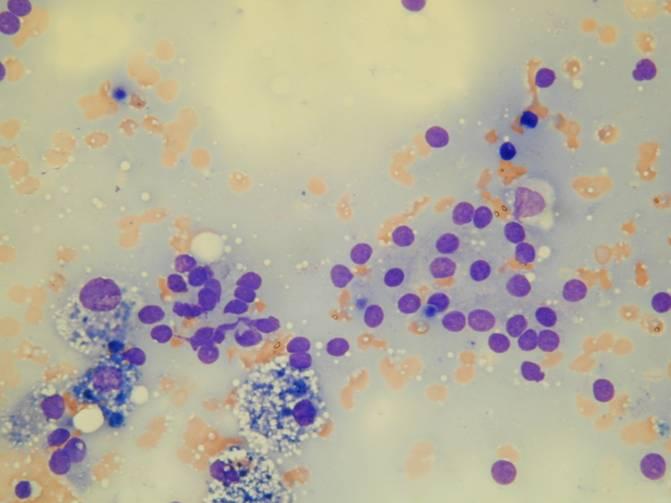
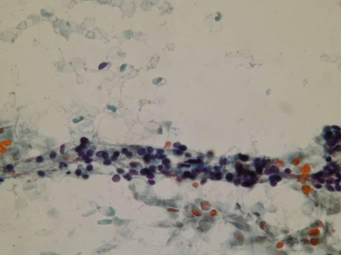
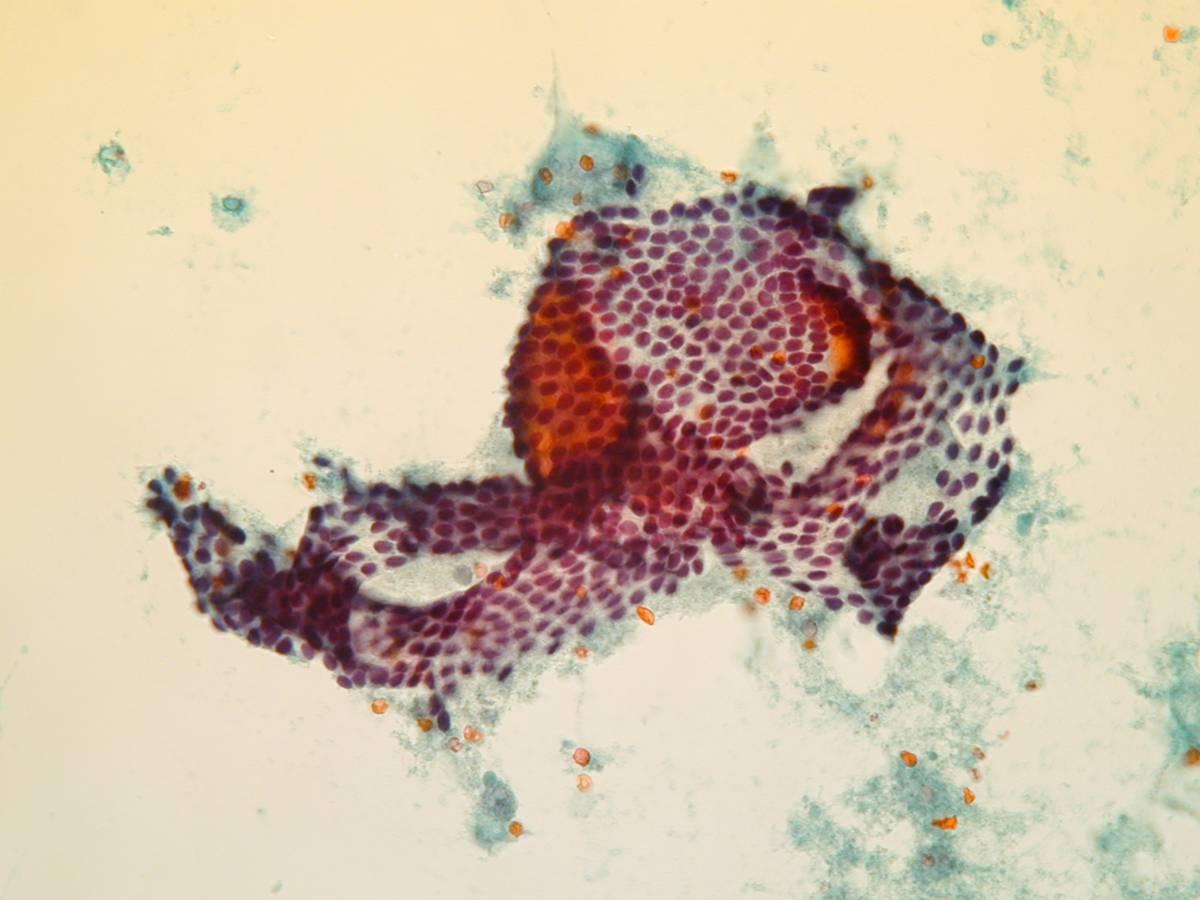
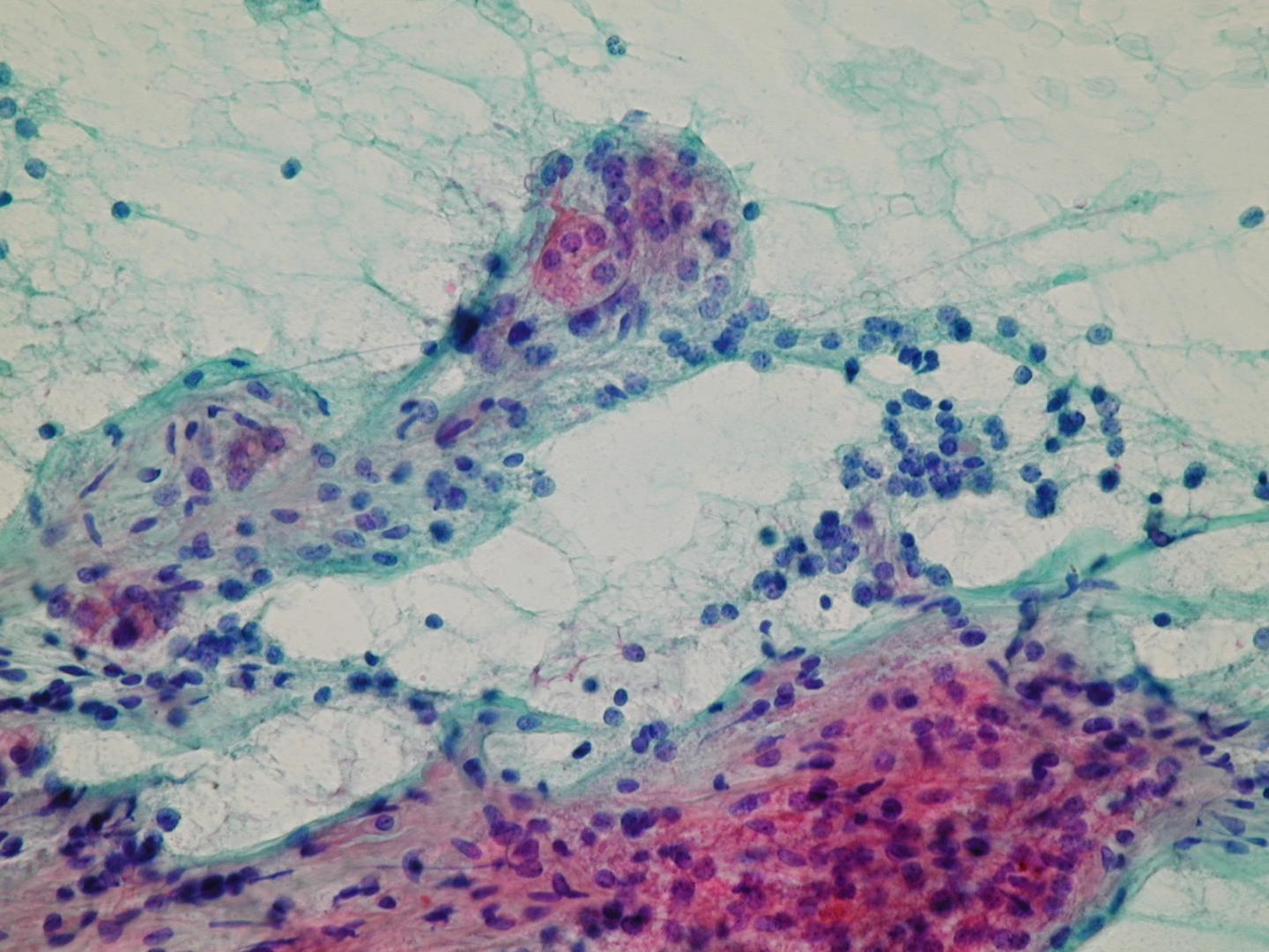
Jest to guzowate powi?kszenie tarczycy, powsta?e w wyniku zak?óce? w produkcji hormonów, które jest zwykle asymetryczne i czasami osi?ga znaczne rozmiary. Komórki p?cherzykowe wykazuj? cechy hiperplazji, doprowadzaj?c do powstania wielu guzków. Guzki, zazwyczaj nieotorebkowane, mog? si? znacz?co ró?ni? w obrazie mikroskopowym: niektóre zbudowane s? z bardzo du?ych p?cherzyków zawieraj?cych koloid, inne s? bardziej bogato komórkowe z niewielk? ilo?ci? koloidu. Rozrost tych guzków prowadzi do wylewów krwotocznych, bliznowacenia, tworzenia ma?ych torbieli i dystroficznych nawapnie?.   

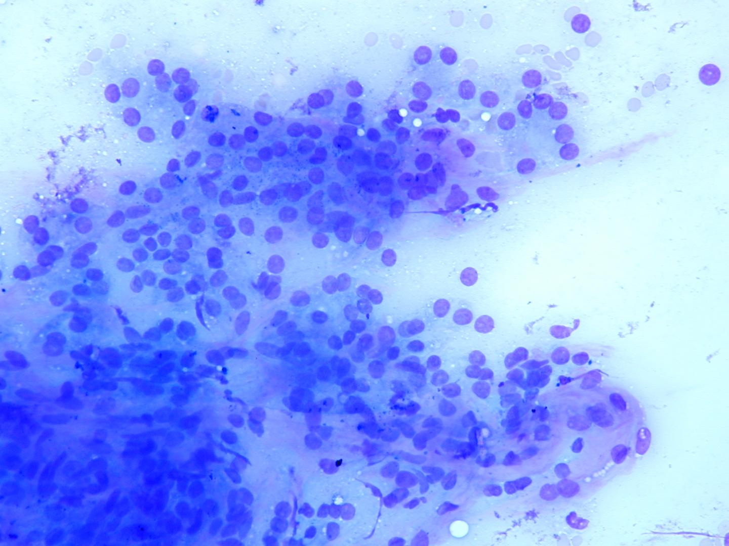
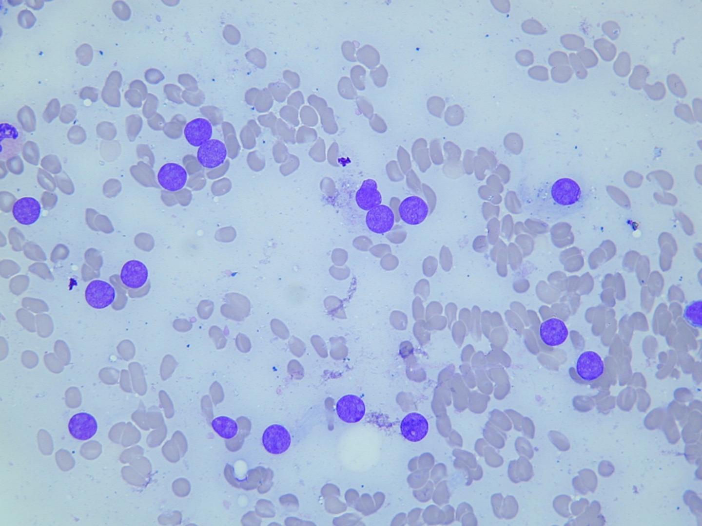
Diagnostyczne cechy cytologiczne

  • ?agodne tyreocyty tworz?ce p?cherzyki lub p?aty
  • obfity p?ynny koloid
  • piankowate makrofagi
Fluid colloid with scattered follicles and histiocytes – Groups of bland thyroid cells in a microfollicular arrangement, bare nuclei of thyroid cells and fluid colloid in the background. A few macrophages are also present. This pattern is associated with benign disease.
Pigmented macrophages and few small epithelial cells which appear hypofuctioning (small, hyperchromatic nuclei with scanty, denser cytoplasm.
Fluid colloid, bland epithelial cells and histiocytes – Pigmented and vacuolated macrophages admixed with bland epithelial cells and bare nuclei. Note the fluid colloid background.
Pigmented and vacuolated macrophages, bland follicular cells – Pigmented macrophages with bland follicular cells and bare nuclei in the background.
A streak of bland follicular cells – A common finding in poorly cellular samples: a columnar group of slightly hyperchromatic but otherwise bland follicular nuclei; these may represent hypofunctioning cells. They may be confused with lymphocytes which usually have a clearly identifiable peripheral rim of cytoplasm.
A folded sheet of bland epithelial cells – These folded larger sheets of bland epithelial cells usually line larger follicles (macrofollicles). Note the monolayered aspect of the cells and regular nuclei. 
Bland follicular cells – Numerous follicles and isolated epithelial cells trapped by stromal septa. Note the elongated hyperchromatic nuclei of fibroblasts in the upper left corner.
3D follicles – A microbiopsy, as may be  obtained with larger needles, in which 3D follicles are separated by collagenous septa; the margins of the follicles are regular. This pattern is usually seen in FNAs from nodular goitres.
Bare nuclei – Numerous bare nuclei, probably derived from functional epithelial cells with abundant, clear and fragile cytoplasm (arrow) which exhibit a round nucleus with open chromatin and visible nucleolus.