This content is also available in:
![]() Español
Español ![]() Čeština
Čeština ![]() Magyar
Magyar ![]() Polski
Polski ![]() Türkçe
Türkçe
Malignant effusions
Some tumours have a greater tendency than others to spread to the serosal cavities. The most common causes of malignant pleural effusion are lung cancer in men and breast cancer in women. In some cases, a malignant pleural effusion can be the first manifestation of lung cancer, whereas it is very uncommon for breast cancer to manifest itself initially as a malignant effusion. The most common occult sources of a peritoneal effusion are intestinal, gastric and pancreatic cancer in men and ovarian cancer in women. The presence of malignant cells in pleura, pericardial or peritoneal fluids is associated with a poor prognosis. In children, the most common cause of a malignant pleural or peritoneal effusion is a non-Hodgkin lymphoma. Primary malignancies of the serosal membranes (malignant mesothelioma) can present as malignant effusion as well.
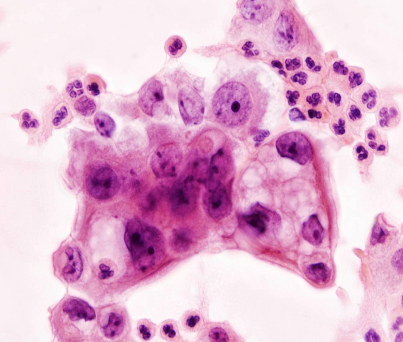
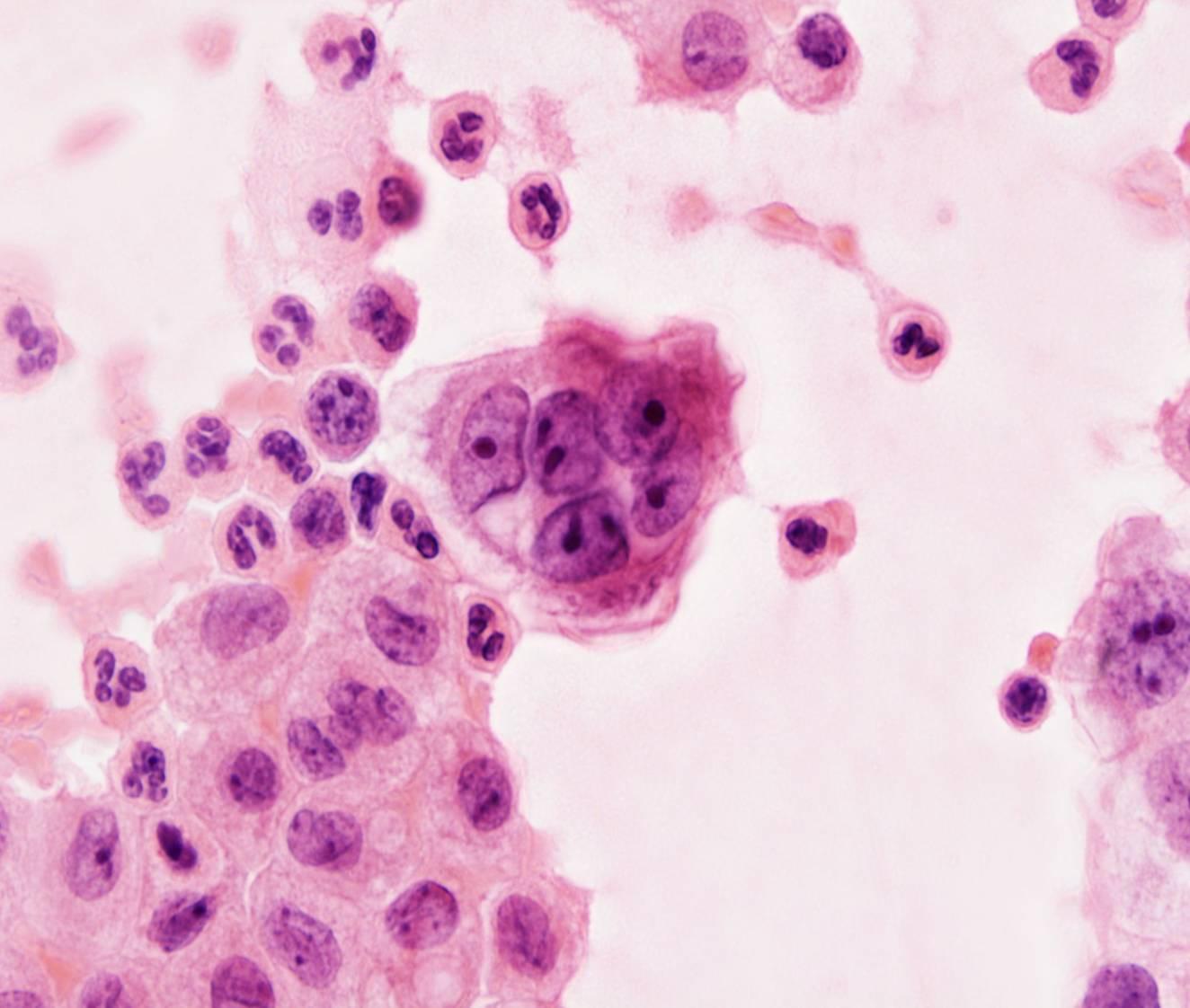
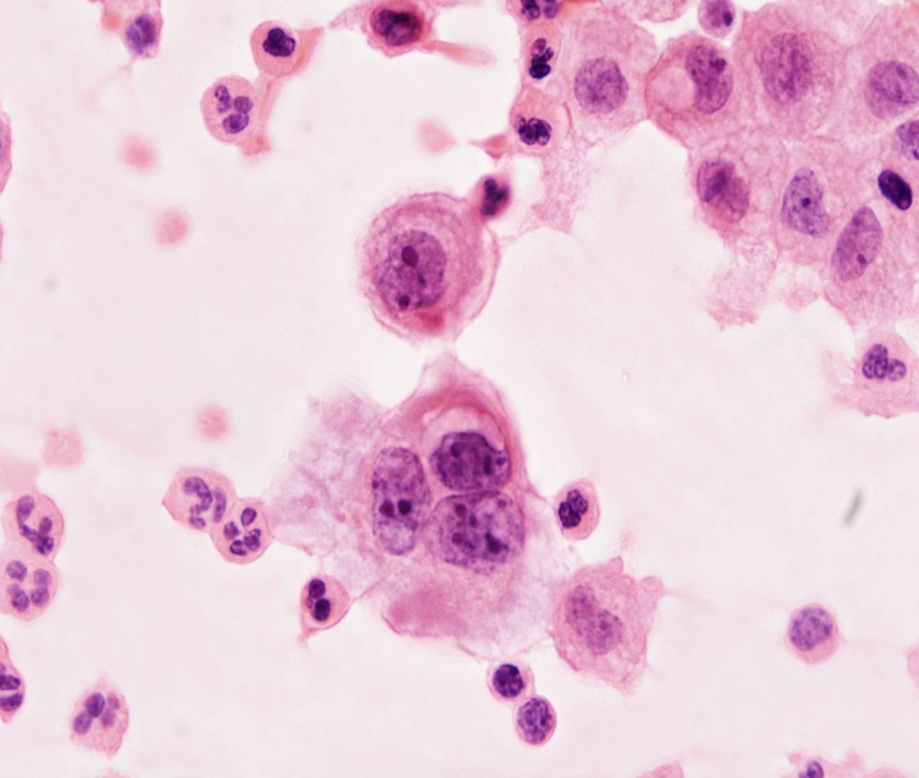
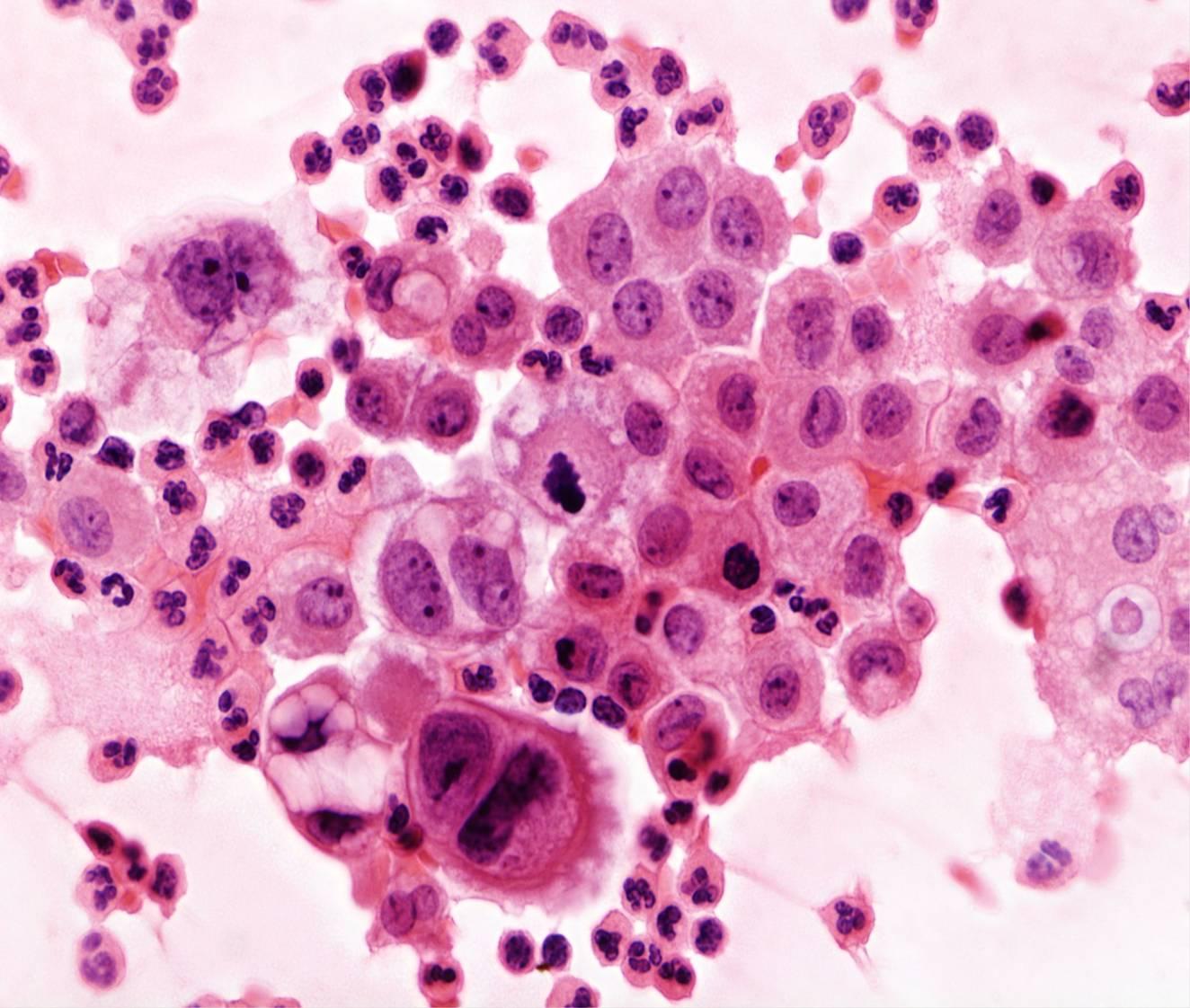
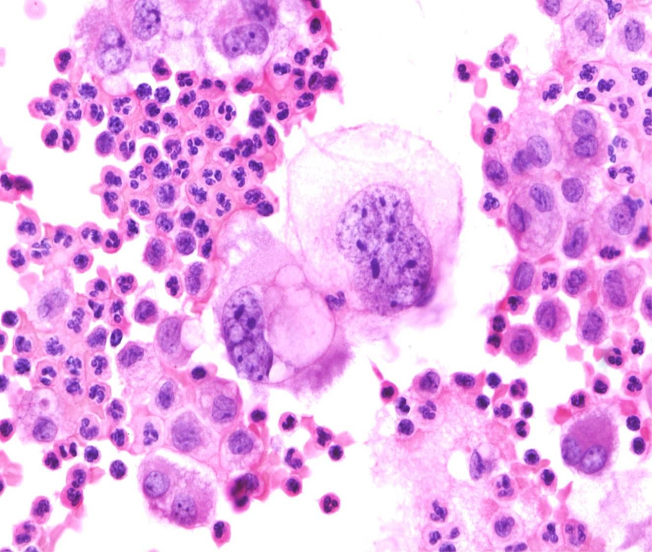
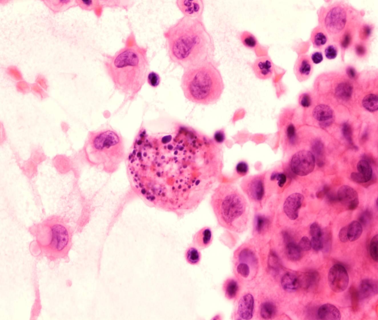
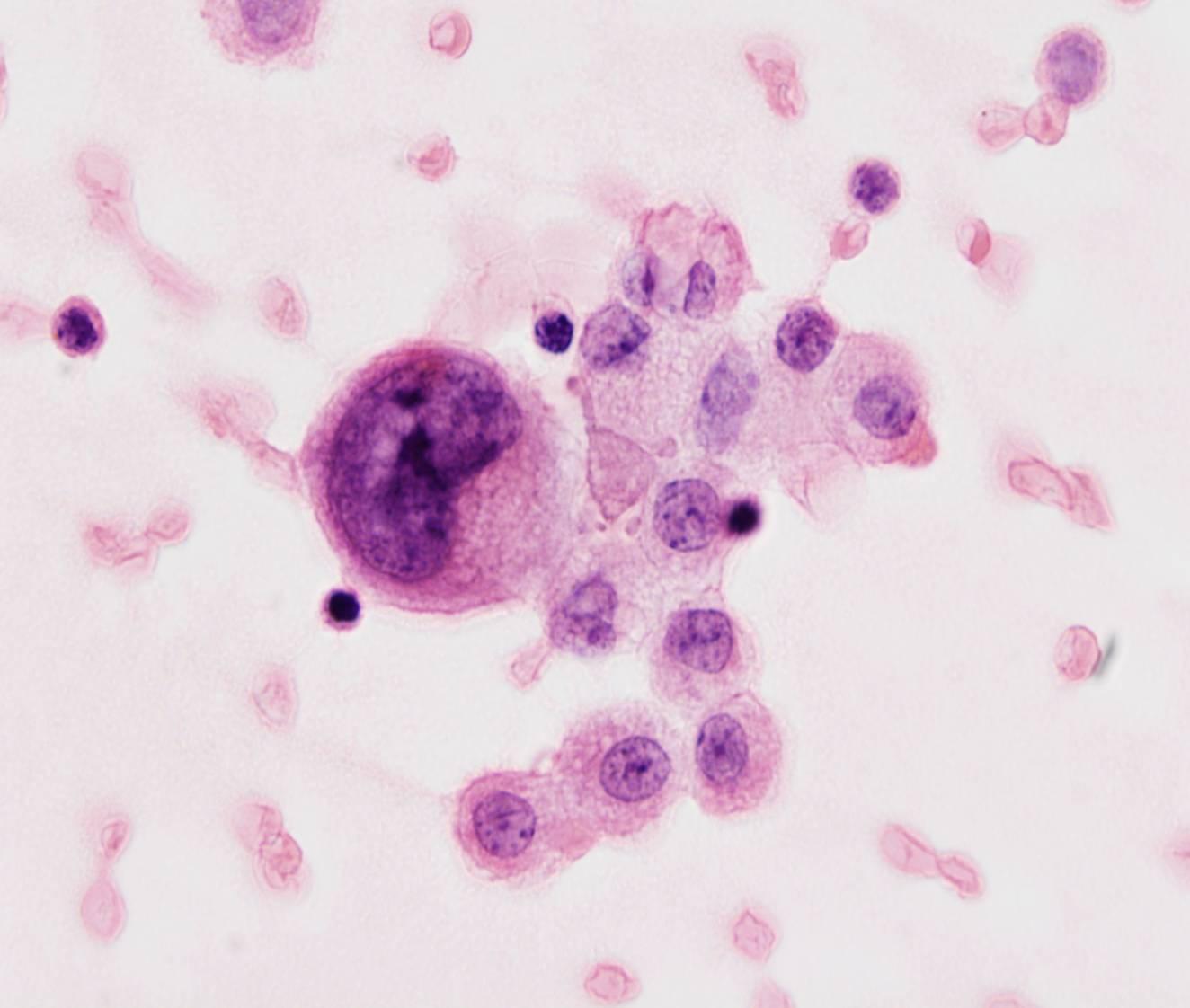
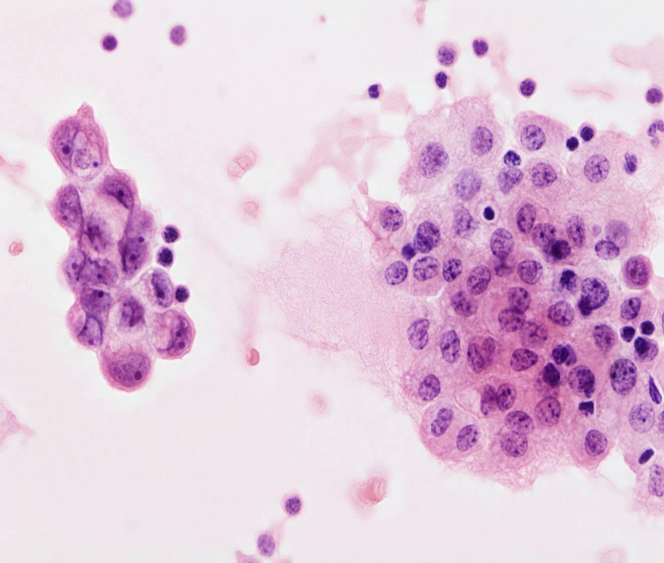
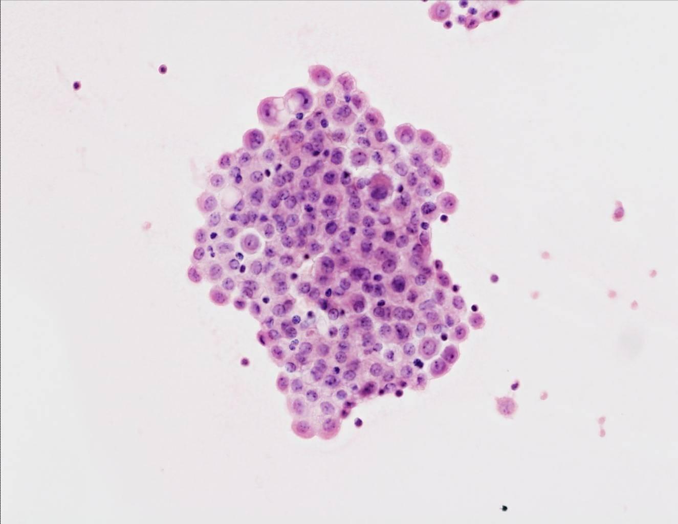
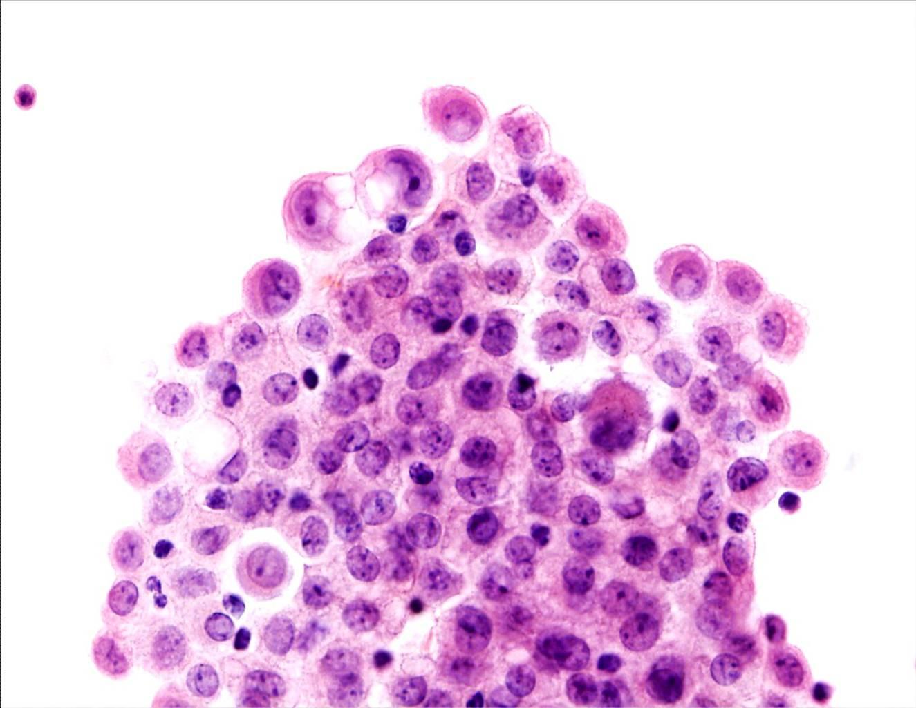
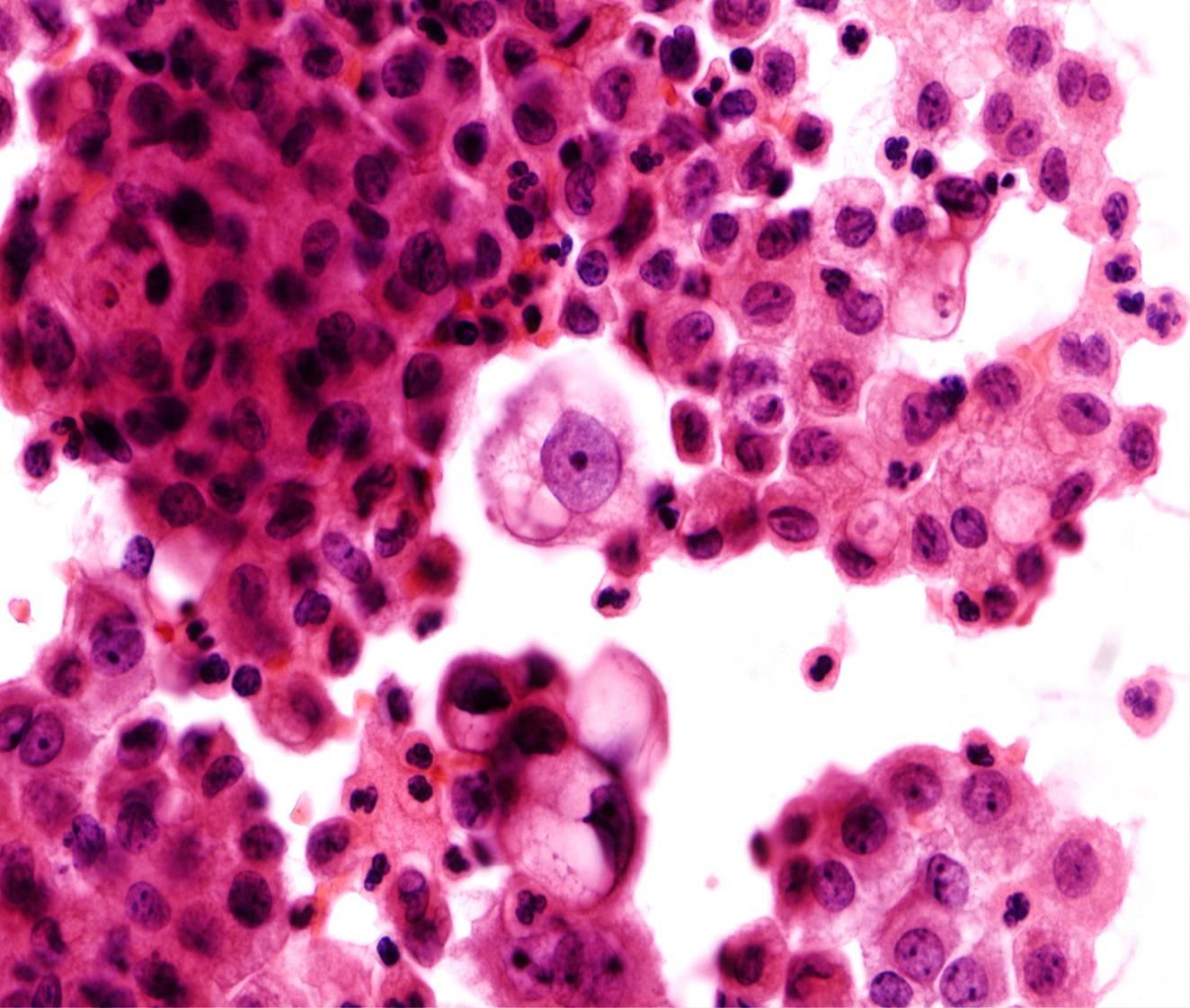
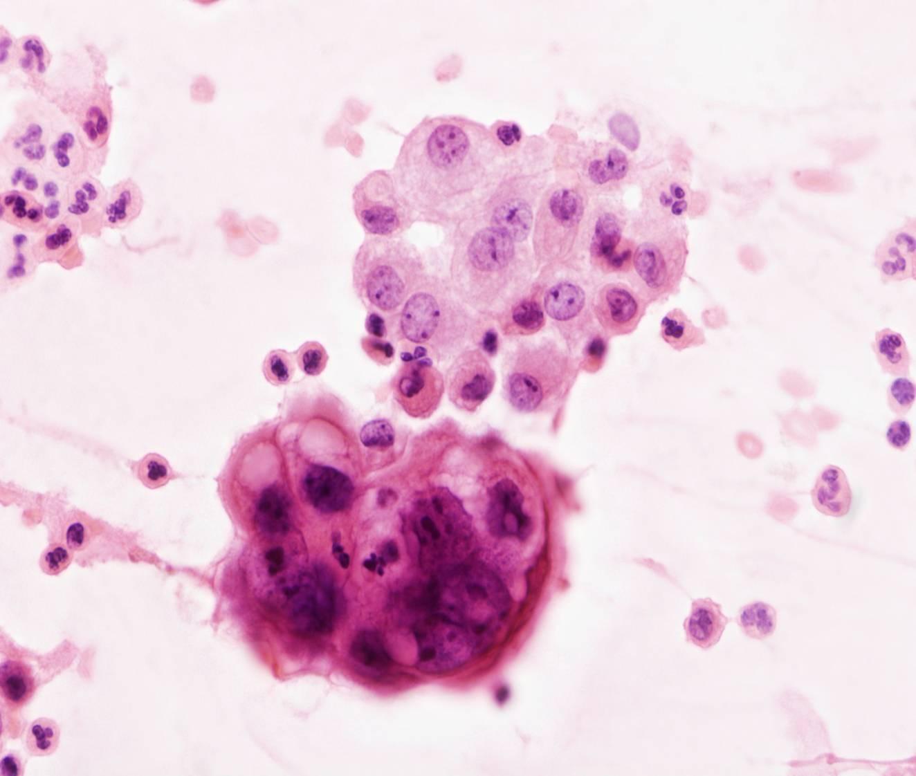
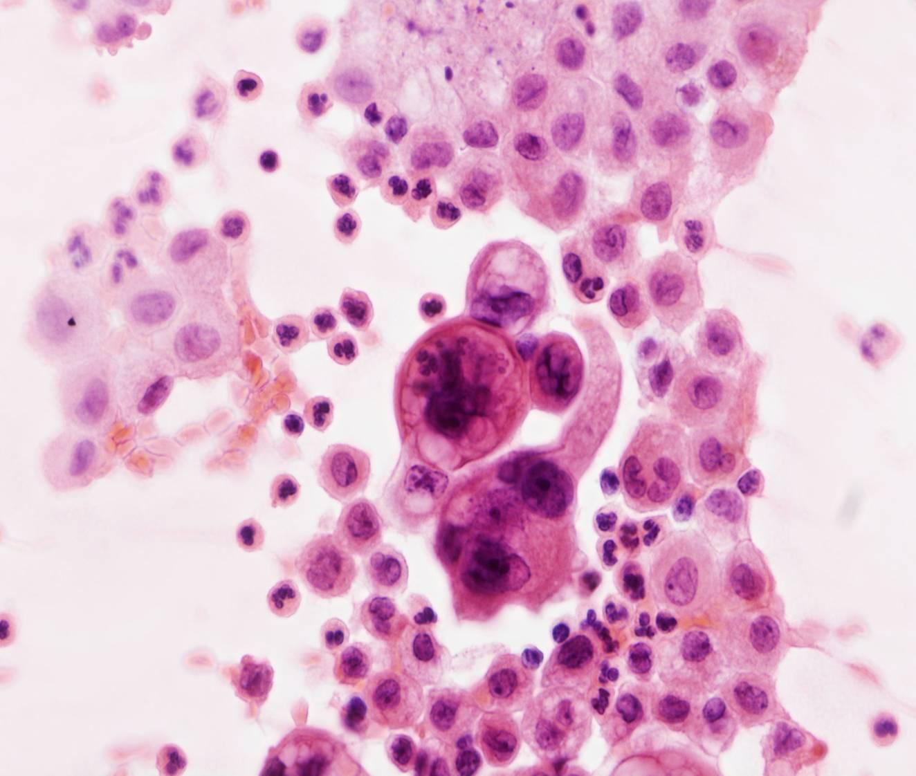
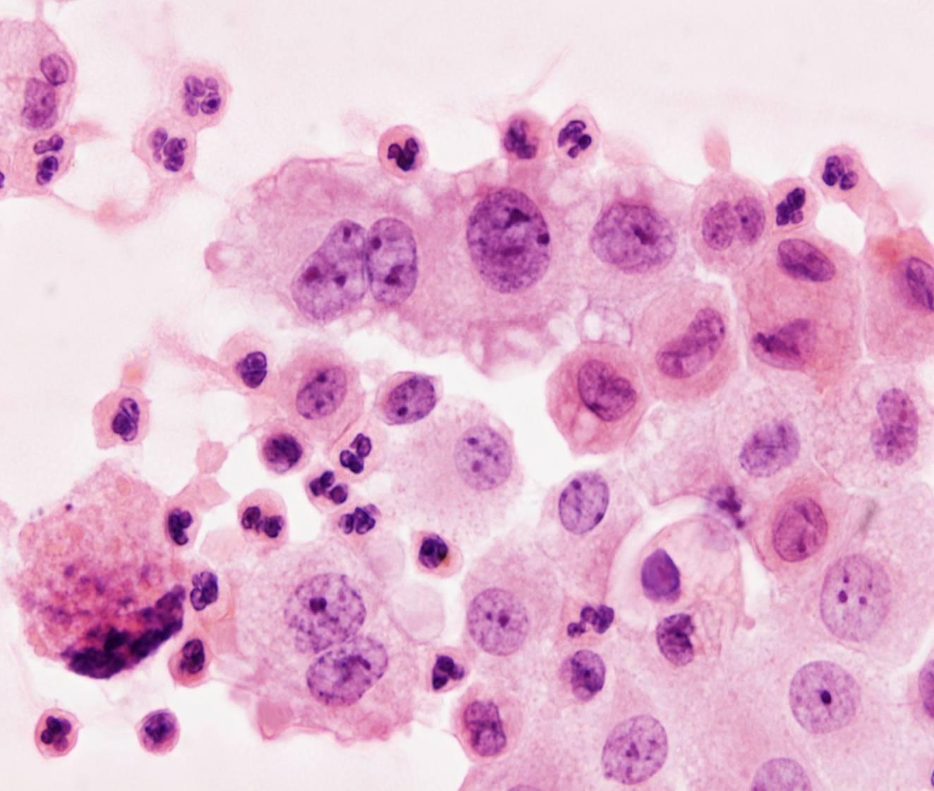
The diagnosis of malignancy rests exclusively on morphologic features. Ancillary methods (ICC, cytochemistry) are only useful for tumour typing.
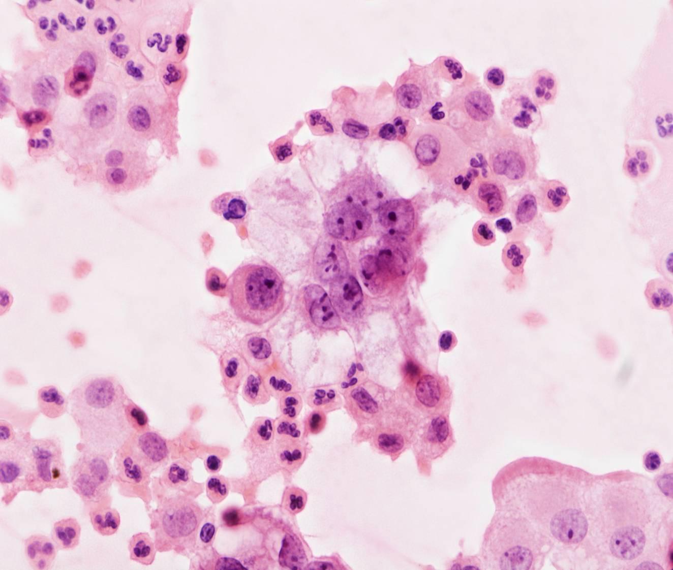
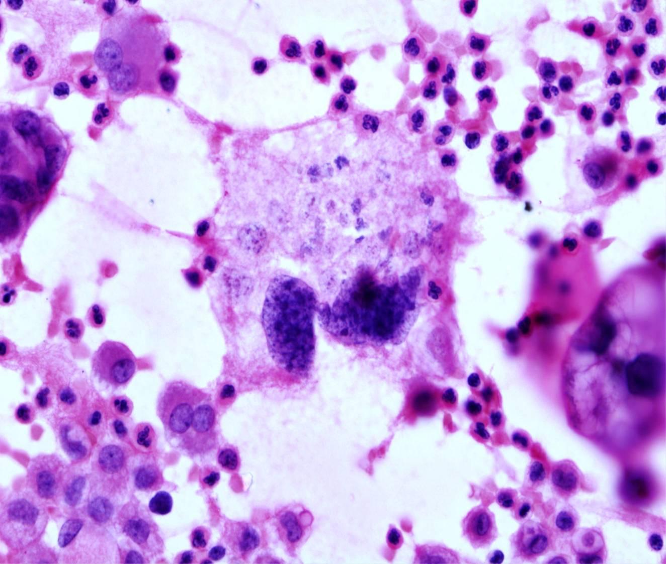
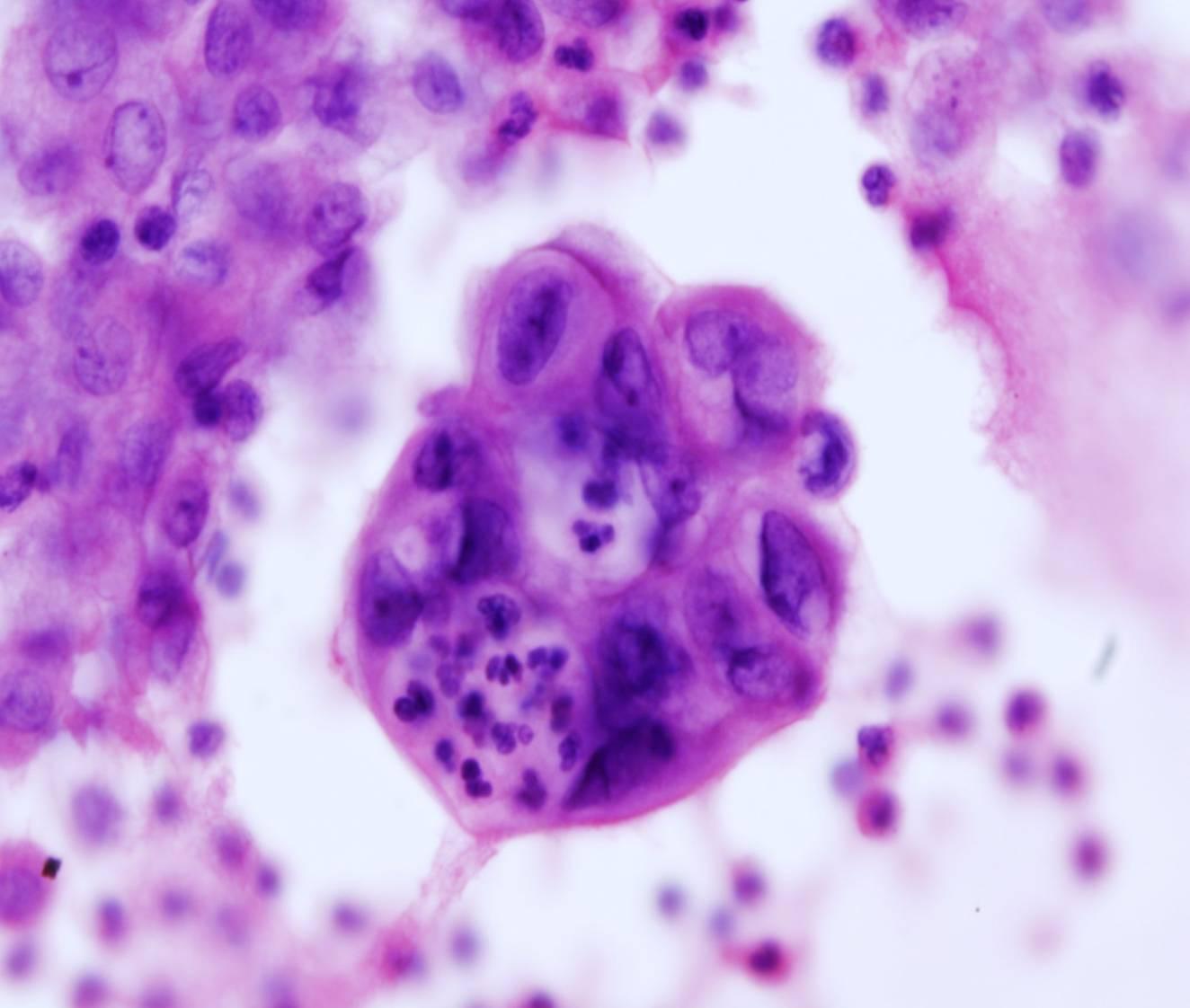
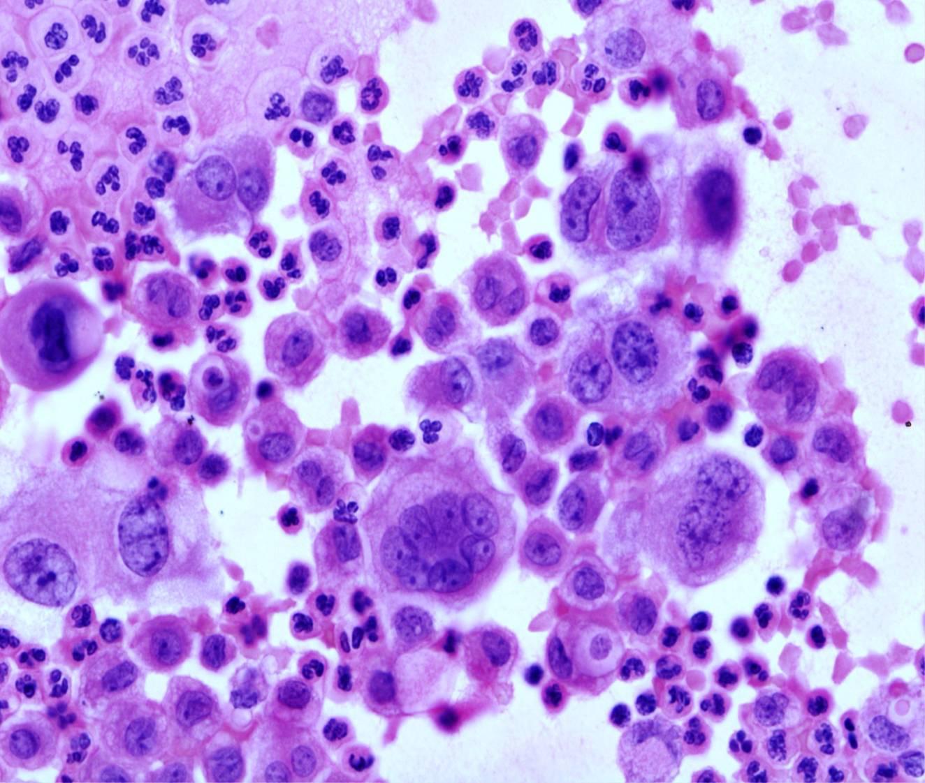
Tips for detecting malignant cells:
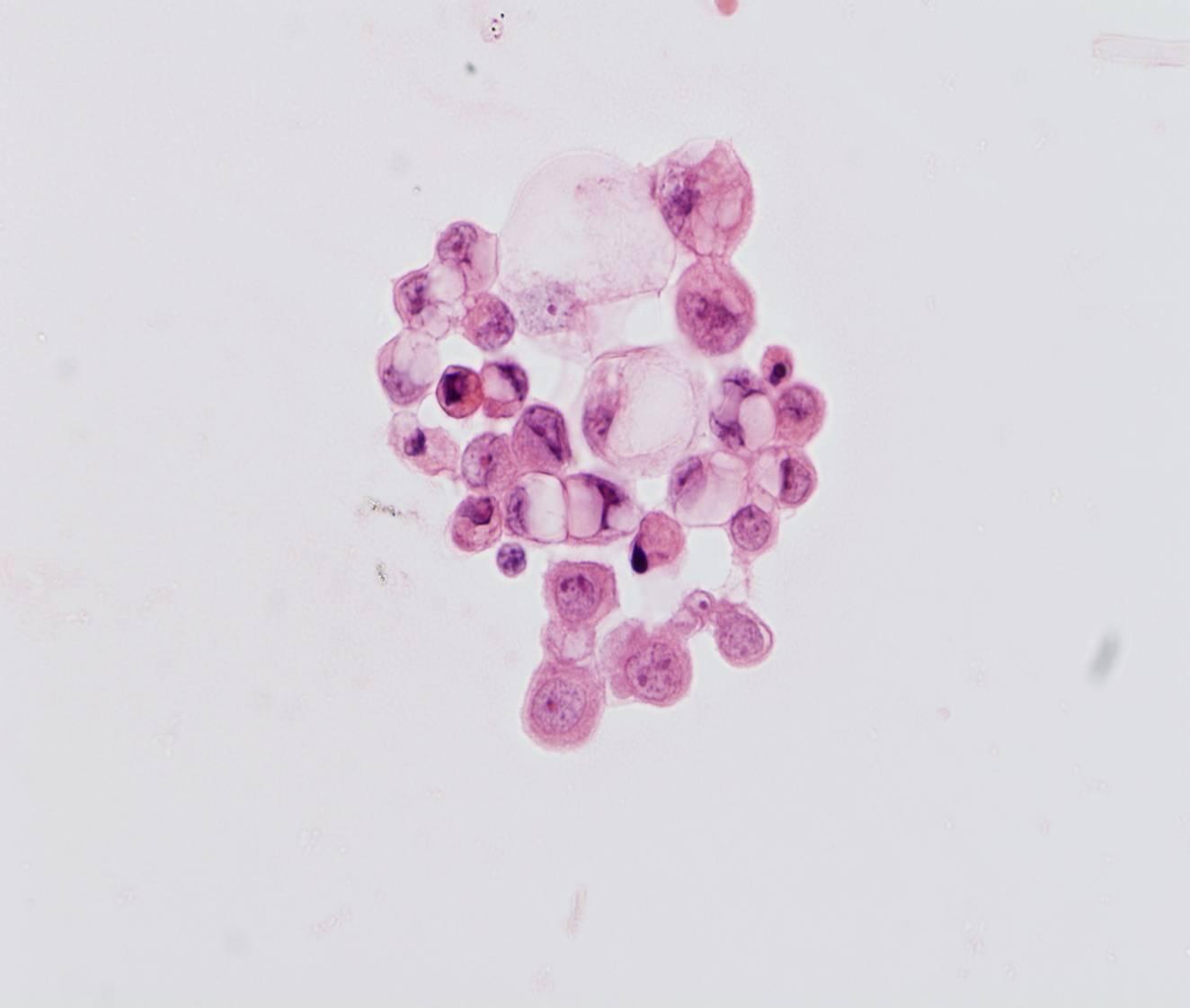
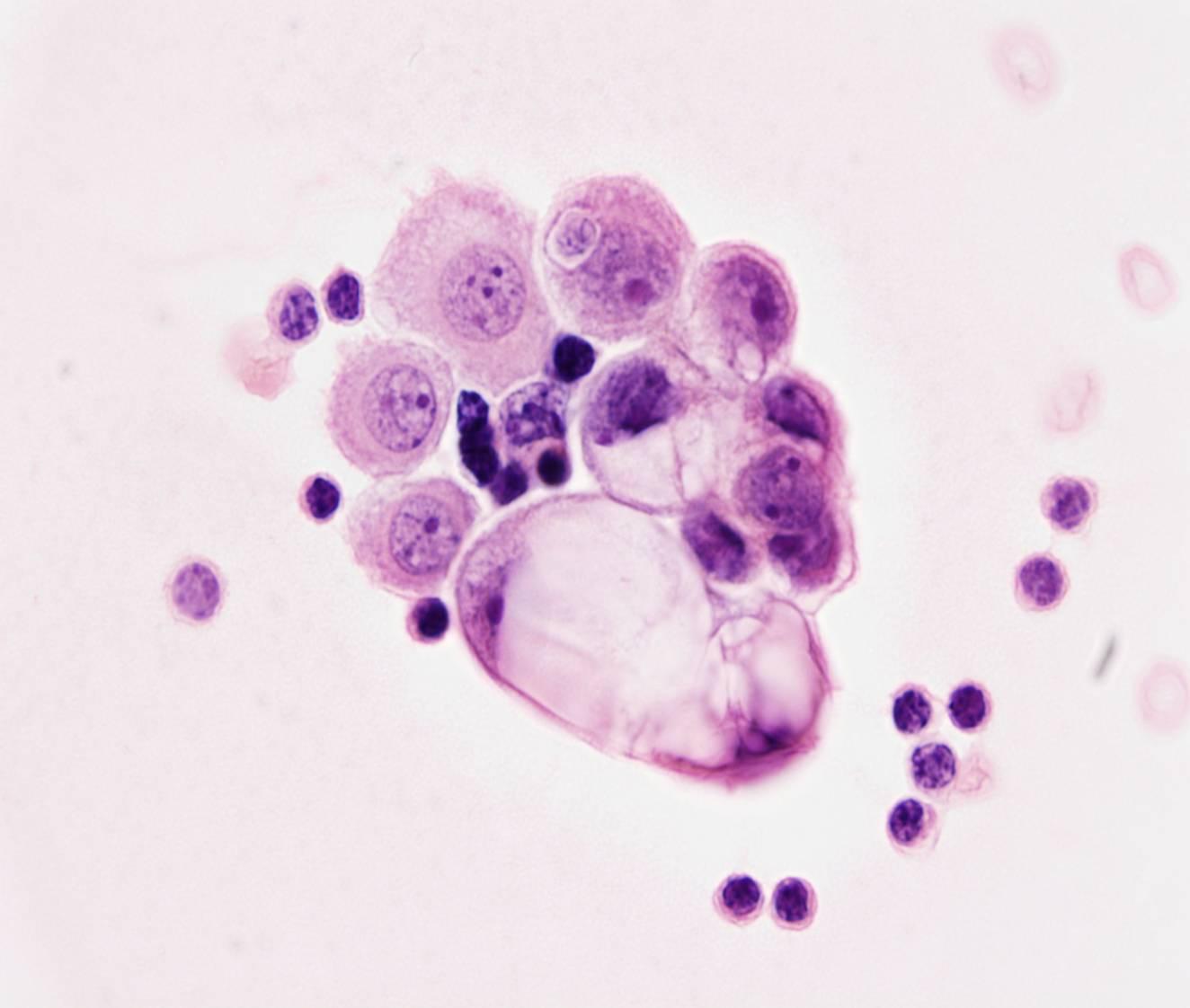
- Second population
- Numerous large clusters
- Lacunae (cell block sections)
After locating some benign mesothelial cells, malignant cells can be identified as a second population of cells that are clearly different. They are not necessarily larger than the mesothelial cells; some are about the same size, with high N/C ratio, nuclear hyperchromasia or macronucleoli. In malignant mesothelioma, however, a sharp distinction between benign and neoplastic mesothelial cells is rarely seen. Normal mesothelial cells virtually never form large tight clusters; samples with numerous cell aggregates are easily recognized as malignant. They must not be confused with loosely clustered cells, which are a common artefact of cytocentrifugation and liquid-based preparation. Malignant clusters are tightly cohesive. In cell block sections malignant cells are often situated in lacunae, which consist of clear spaces surrounding individual cells or groups of cells. However, lacunae can also be seen in many benign effusions.