logo efcs bianco

Cytologically benign follicular lesions

This content is also available in: Español Čeština Magyar Polski

Bookmark (0)

No account yet? Register

Cytologic diagnostic features

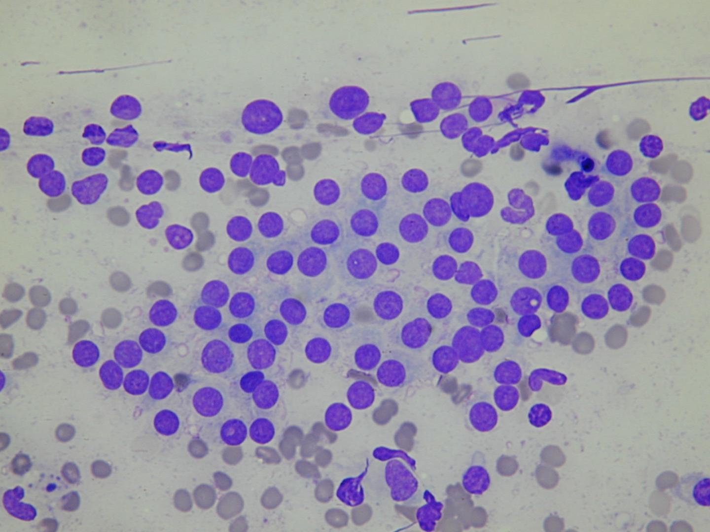
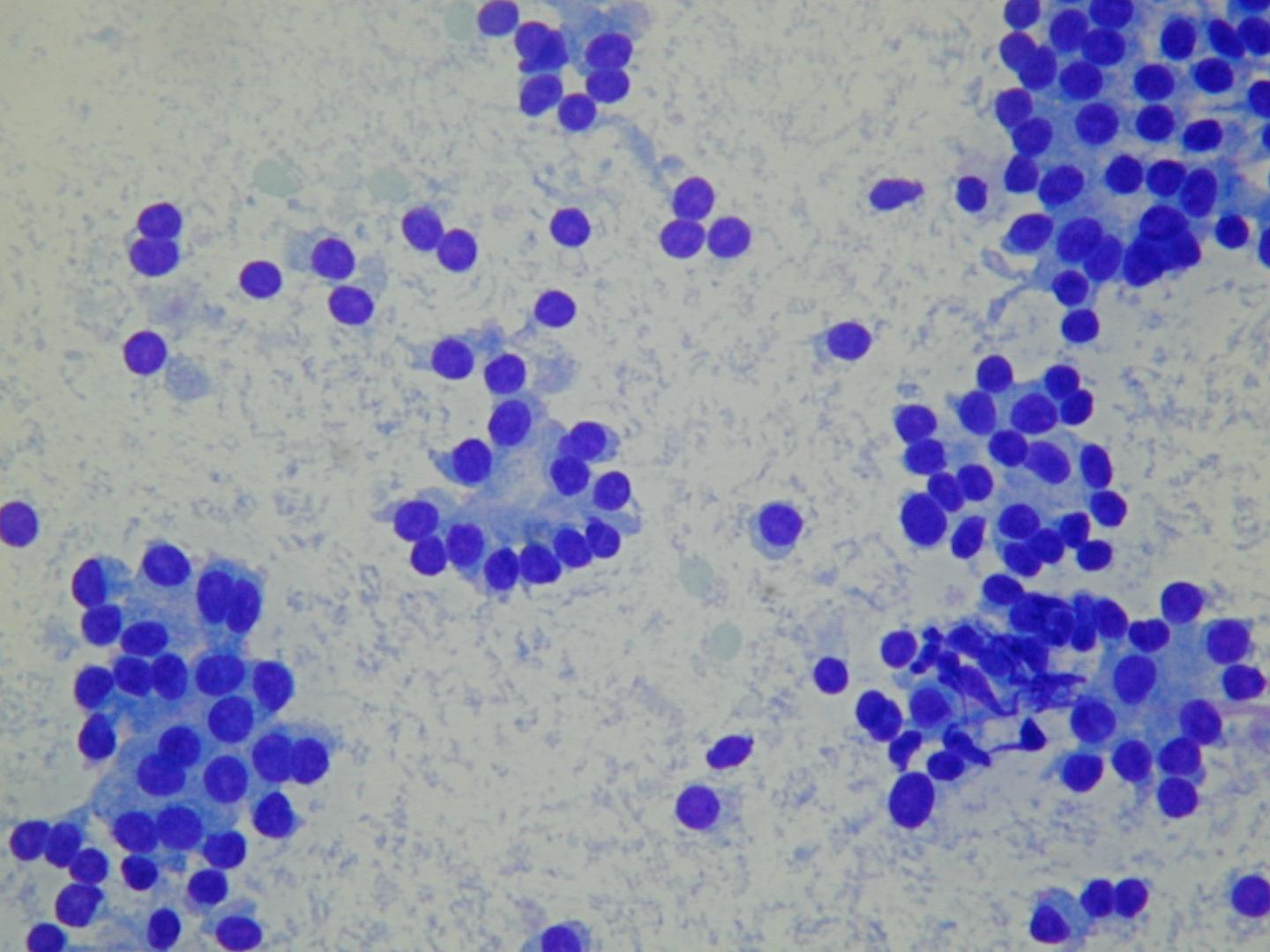
  • low or moderate cellularity
  • cohesive cells
  • predominantly microfollicular pattern
  • uniform, evenly spaced follicular cells
  • round nucleus, finely granular chromatin
  • scanty or moderate cytoplasm
  • few macrophages
  • bare nuclei

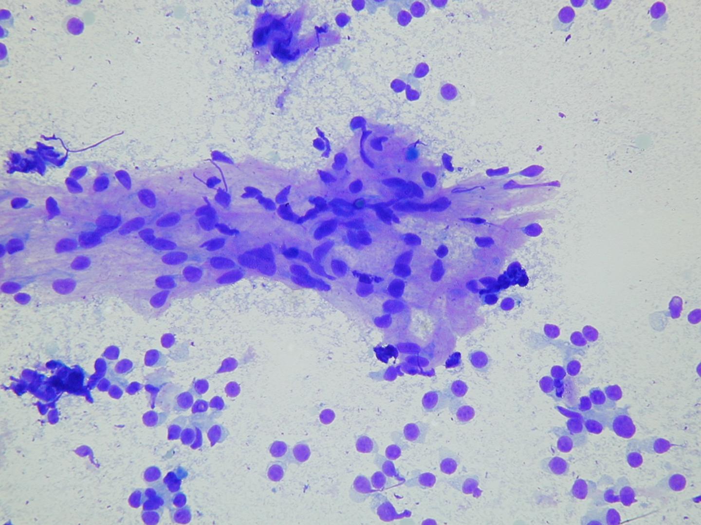
Colloid is usually abundant, appearing as amorphous blobs or as a thin translucent film with bubbles and linear cracks.

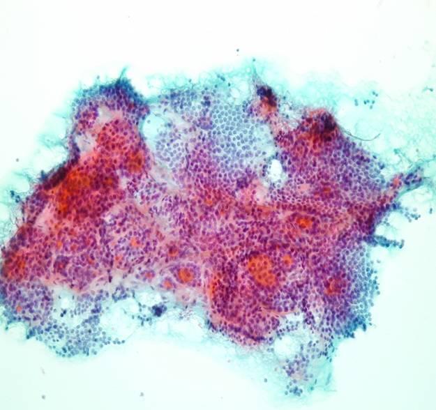
Some benign follicular lesions are hypercellular and even focal cytologic atypia may occur. Occasional large, spindle-shaped cells may be present, representing either reactive stromal elements or altered follicular cells lining areas of cystic degeneration. Focal Hürthle cell change may also occur. If microfollicles are few and atypia is focal, a diagnosis of cytologically benign nodule should be made, even if the specimen is cellular. Patients with this diagnosis have to be followed up at an appropriate interval. 

A microbiopsy from a nodular goitre. A thick fragment in which round 3D follicles are separated by collagenous stroma. Orangiophilic colloid is visible inside some follicles whose contours are round. Where thinner a monolayer arrangement can be identified. 
Bland thyrocytes in a microfollicular arrangement. A monolayered sheet of bland thyrocytes with bland chromatin and moderate amount of pale, delicate cytoplasm. In the upper corner a follicle lined by similar cells is observed.  
Bare nuclei and microfollicles. Few microfollicles lined by uniform cells with delicate cytoplasm, round nuclei with fine chromatin and surrounded by bare nuclei (MGG).
A very low power view of a cellular lesion. A very lower power view of a MGG stained smear from a follicular lesion. It is very cellular, colloid is not visible and the cells are arranged in sheets, follicles or lie singly. At this magnification no commitment can be made as whether it is benign, suspicious or malignant. 
At high power the cells are uniform. A microfollicular pattern of regular, uniform thyroid cells exhibiting some “flaring” at their surface, usually seen in functioning cells; they are also monolayered and no reowding is observed.
A trabecular arrangement. The same cells may exhibit a trabecular arrangement; in this case the minimal degree of anisonucleosis is well within normal limits. Anisonucleosis (variability in the volume but not the shape of nuclei) is of very little significance in all endocrine glands and is certainly not a criteria for malignancy (MGG).
Bland, less cohesive thyrocytes. Nuclear detail is better seen in Papanicolaou stain: chromatin is fine and small chromocentres can be seen. Nuclear membrane is regular and thin while cytoplasm is delicate and transparent.
A cellular aspirate at low power. A medium power view of a microfollicular adenoma (MGG). At this magnification lack of background colloid, uniformity of cells and microfollicular pattern are obvious. In the absence of nuclear atypia, crowding or papillary architecture these lesions are classified differently either as follicular lesion, cytologically benign, Thy3 or follicular lesion – indeterminate. In all cases the physician must know that criteria to predict a well differentiated follicular carcinoma are only histological following excision of the lesion to search for capsular or vascular invasion. However an aggressive surgical approach will result in many benign lesions being excised.  
The cells are uniform and arranged in follicles. The same lesion at higher power confirms lack of nuclear atypia and a dominant microfollicular pattern. Bare nuclei are seen in the background, a feature which is reassuring as it is usually associated with benign lesions. 

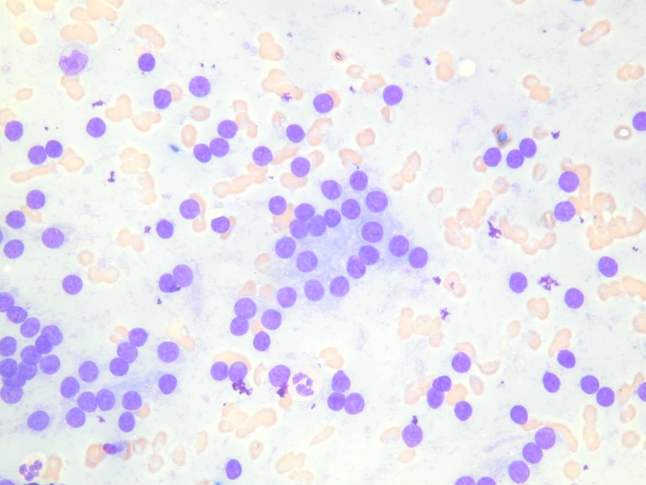
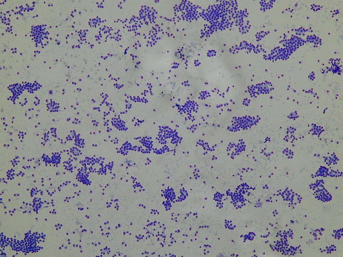
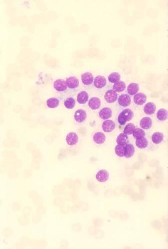
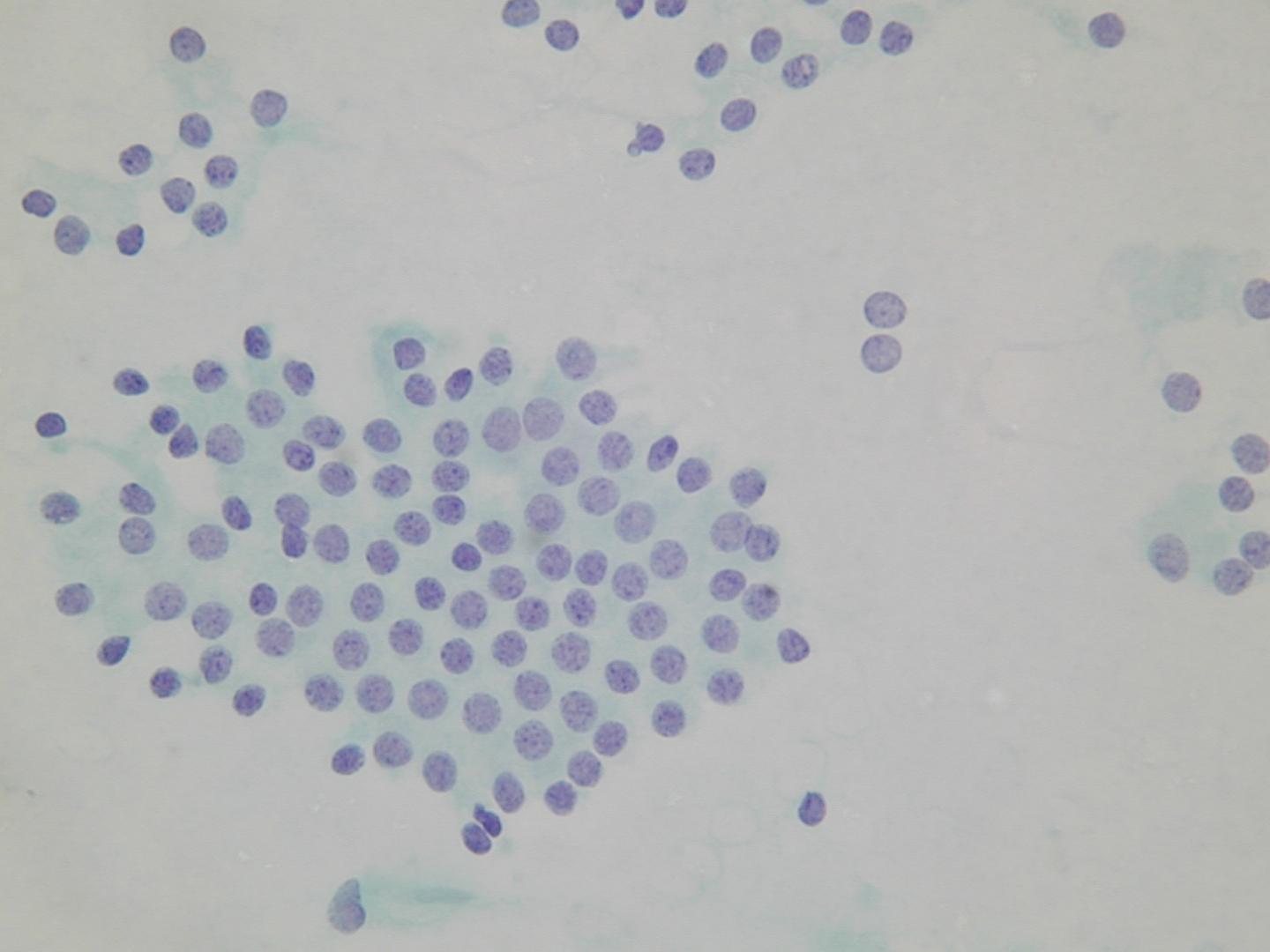
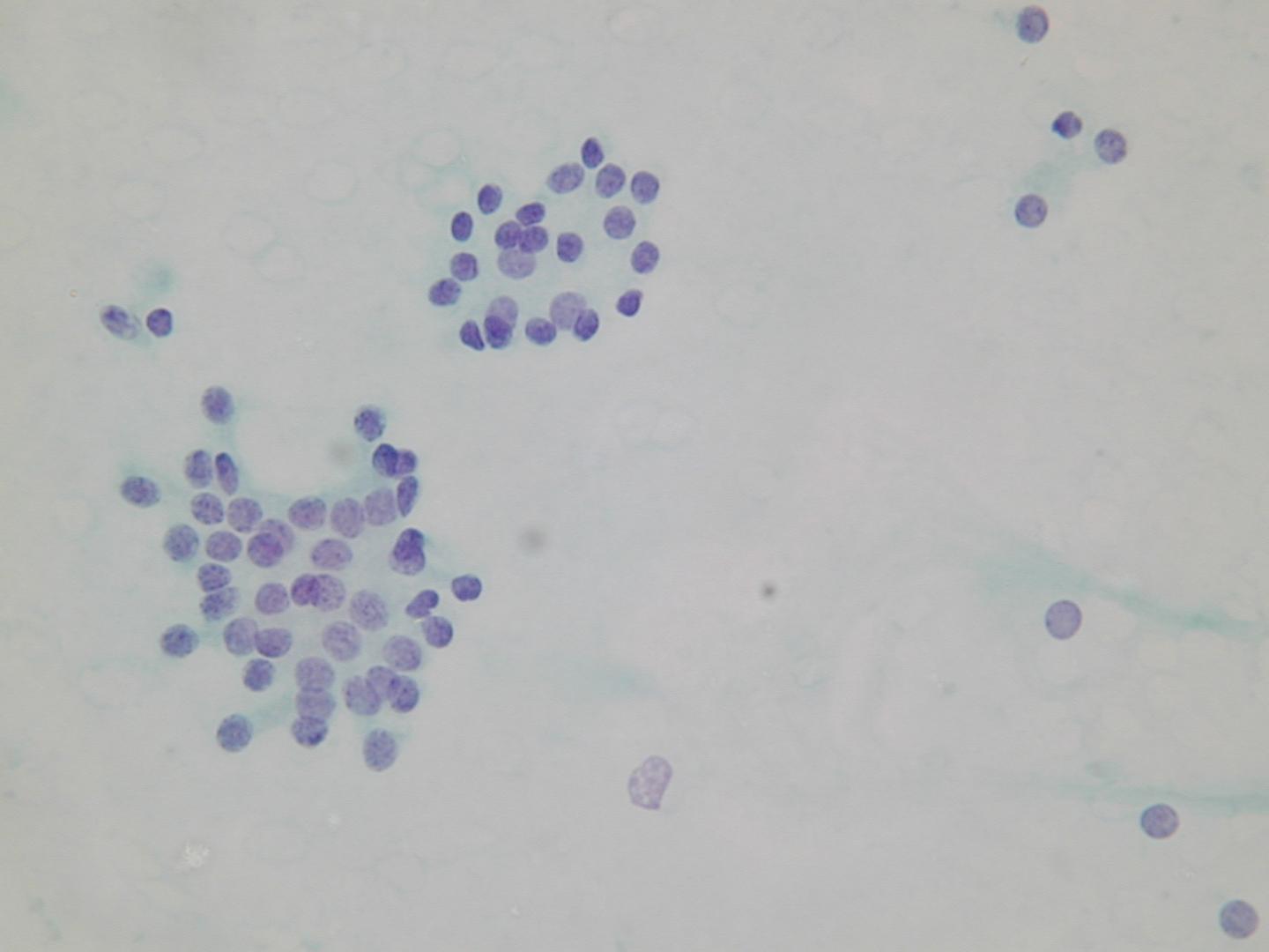
A microfollicular pattern:
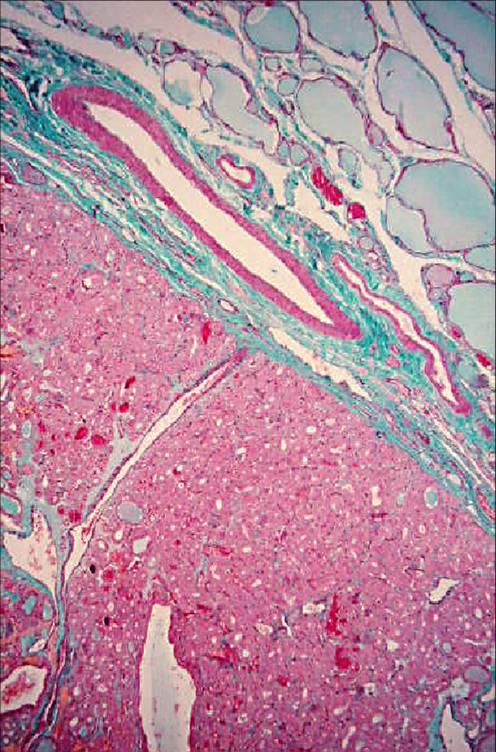
A microfollicular pattern. A microfollicular pattern (MGG); cells appear uniform and no crowding is observed, however few discohesive cells maintain their cytoplasm. 
A stromal fragment with mesenchymal nuclei. At MGG stroma is pinkish and fibrillary with mesenchymal cells appearing as elongated and usually hyperchromatic nuclei; the thyroid cells maintain a follicular architecture and are not arranged perpendicularly to the stroma as expected in a papillary lesion. 
A monolayered sheet of bland thyrocytes. A sheet of monoyared bland thyrocytes surrounded by smaller follicles and bare nuclei. 
A microfollicular arrangement. Bland follicles and bare nuclei. 

Follicular adenoma

It is a benign neoplasm, presenting as a single nodule, usually not greater than 3 cm in diameter. Some of them can produce thyroid hormones and consequently cause hyperthyroidism (functioning or ‘hot’ adenomas). The hystologic pattern may vary: macrofollicular (composed of large follicles filled with colloid), microfollicular (with smaller follicles), trabecular (with follicular cells arranged in ribbons).

Classification (no prognostic significance):

  • simple
  • microfollicular
  • trabecular
  • oxyphil
  • atypicalpapillary
  • signet ring cell
Nodular hyperplasia Follicular neoplasia
multiple solitary
poorly encapsulated encapsulated
architectural heterogeneity uniformity of the architecture
cytologic heterogeneity cytologic homogeneity
comparable areas in adjacent gland different from surrounding gland
no compression of surrounding gland compression of surrounding gland

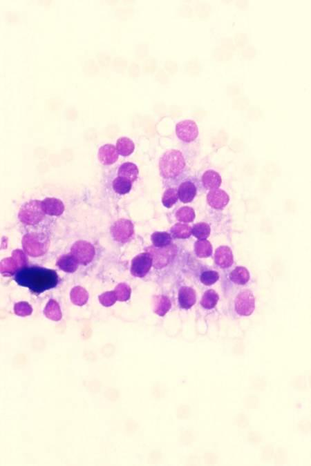
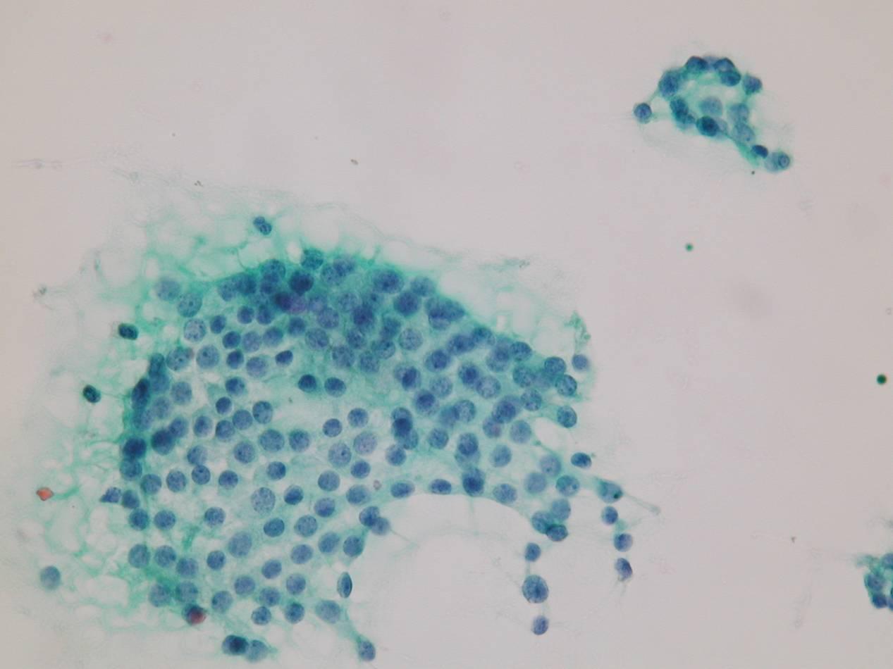
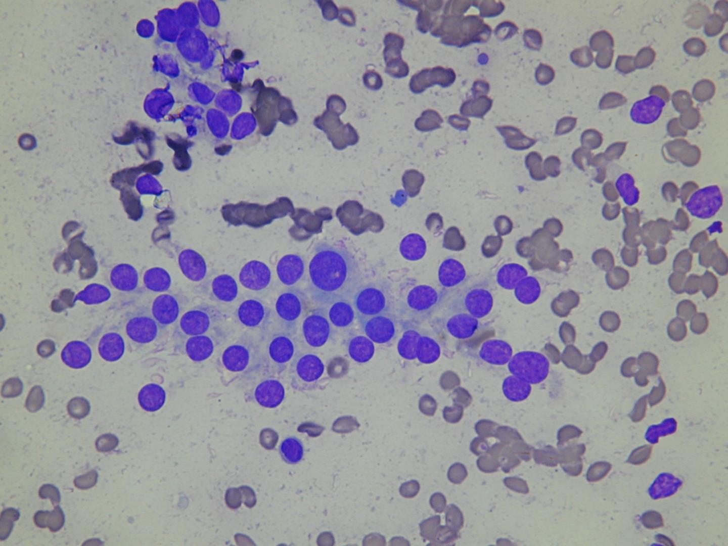
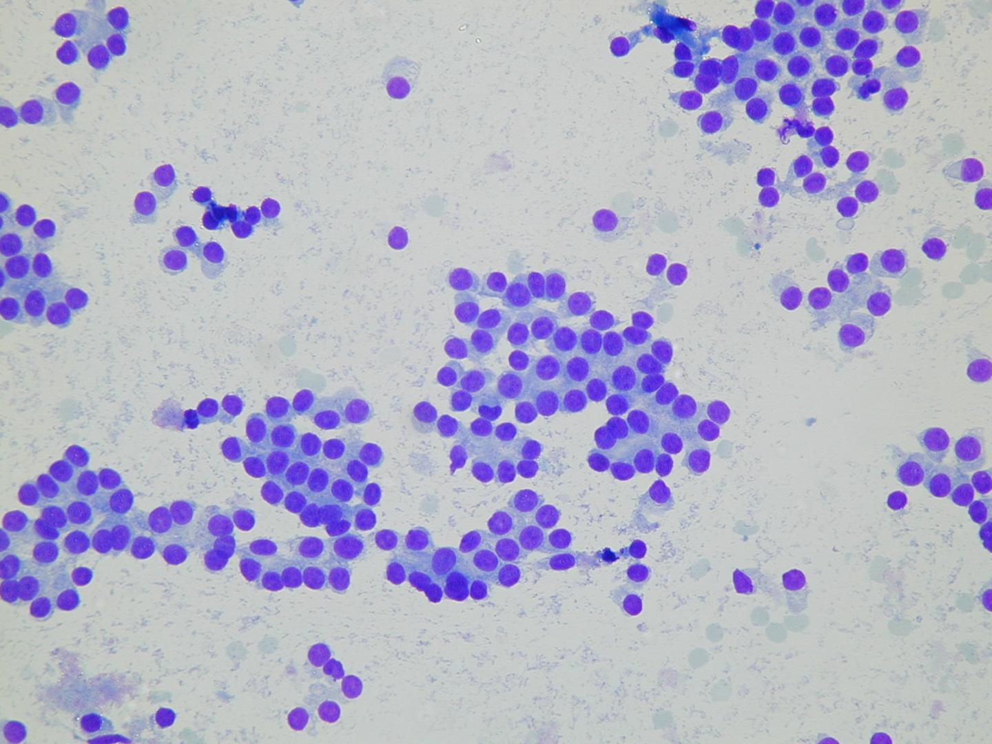
Microfollicular groups:

Microfollicular groups – These microfollicles are lined by thyrocytes showing a round nucleus with ‘open’ chromatin, abundant clear delicate cytoplasm with ‘flares’. These findings are in keeping with functioning cells. 
Microfollicular groups – This follicular aggregate shows uniform, bland thyrocytes with delicate, clear cytoplasm but no ‘flares’. There is no cytological atypia. A benign follicular lesion may be suggested if the colloid/cells ratio is low and bare nuclei are present in the background. 
Microfollicular adenoma (histology) – A low power view of a histologic section of an adenoma which is clearly separated from the surrounding thyroid tissue (Masson Trichrome stain).