This content is also available in:
![]() Español
Español ![]() Čeština
Čeština ![]() Polski
Polski ![]() Türkçe
Türkçe
Benign clear cell tumor (‘sugar’ tumor)
It is a rare tumor, occurring in any age as an asymptomatic peripheral nodule. Because of their immunohistochemical and ultrastructural features, they have been supposed to origin from pericytes; anyway, their origin is a matter of discussion. They consist of polygonal cells with clear, glycogen-rich cytoplasm.
Cytologic diagnostic features
- Clusters of polygonal and spindle-shaped cells
- Vacuolated or granular cytoplasm
- PAS-positive cytoplasm
Differential diagnosis
- Non-small cell carcinoma with clear cell change
- Carcinoid tumor
- Metastatic renal cell carcinoma
Immunocytochemistry is helpful: most sugar tumors are positive for HMB-45, CD34 and S-100 and negative for cytokeratins and CEA.
Pulmonary hamartoma
It presents as an incidental nodule on radiographs, particularly in elderly men. A hamartoma is a neoplasm in an organ that is composed of tissue elements normally found at that site, but growing in a haphazard mass.
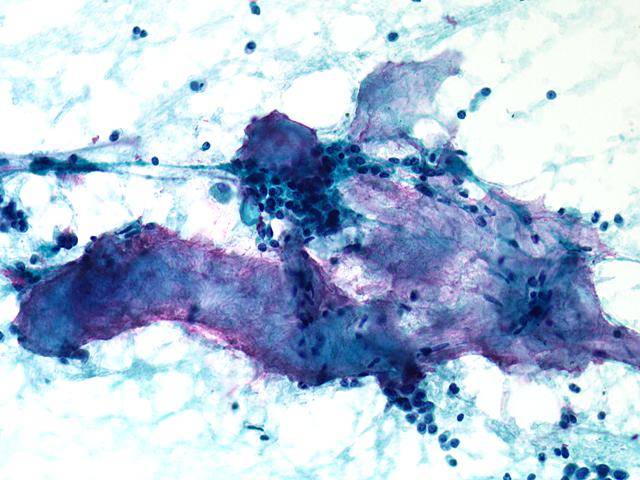
Transthoracic FNA is very sensitive and specific in diagnosing this lesion. A mixture of mesenchymal (mainly fibromyxoid and cartilaginous material) and epithelial elements is seen in FNA material. Bland spindle cells in a fibromyxoid stroma and mature cartilage, with chondrocyte in lacunae, are observed; epithelial glandular cells and adipocytes are often present.
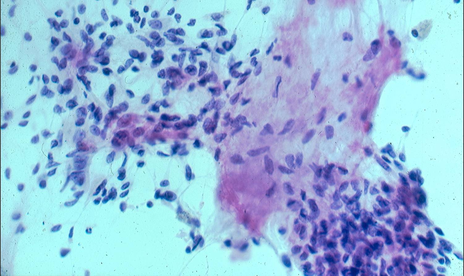
Pulmonary hamartoma.
Note the biphasic morphology with a fragment of benign-appearing epithelium and fibromyxoid stromal tissue.
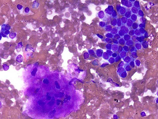
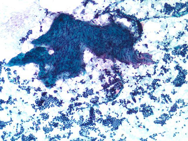
Pulmonary hamartoma.
Biphasic appearance of the tumor with predominance of chondromyxoid stromal tissueand some epithelial cells seen singly and in loose clusters.

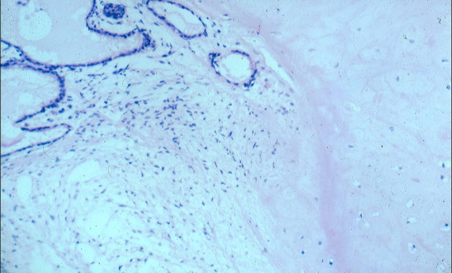
Pulmonary hamartoma.
Epithelial component of the tumor appearing as monolayered fragments of monotonous benign tissue.