Images first published: Glinski et al. Single slide assessment: A highly effective cytological rapid on?site evaluation technique for endobronchial and endoscopic ultrasound?guided fine needle aspiration. Cytopathology 30, 164–172 (2019).
ROSE SSA is utilised for solid lesions in the pancreas. If mucin is present in the adequacy assessed pancreatic preparation or there is a clinical suspicion of a cystic lesion, send a minimum of 2mls of aspirated material (not diluted in saline or cytolyt solution) to clinical chemistry for CEA, Amylase and CA19.9. Check that the clinician has requested the chemistry tests on the request form prior to sending to the laboratory. Please be aware that contamination may occur and intestinal (if transduodenal) or gastic (if trans gastric) epithelium as well as mesothelium or even adrenal or liver cells may be present in the smear.
Diagnostic pitfalls and considerations
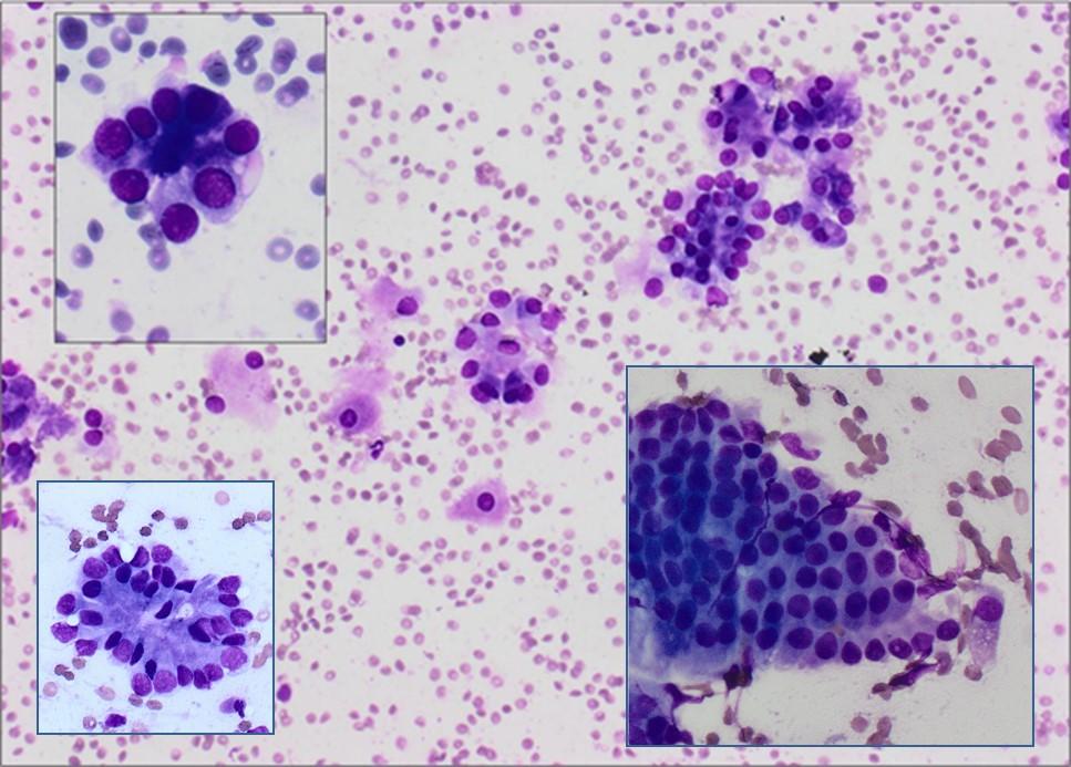
- Ensure both ductal epithelial cells and acinar cells are present for representative pancreatic sampling
- Contaminant sampling. i.e. Gastric epithelial cells
- Necrosis / pancreatitis
- Process all of the residual material in the laboratory even if the ROSE SSA is inadequate. Subsequent diagnostic material can be found in the induced clot of the residual material. The presence of these diagnostic cells had a significant impact on the treatment pathway for these patients. Particularly evident in metastatic renal cell carcinoma where there can be an excess of blood due to the nature of the tumour.
| Adequacy criteria guidance | Requirement |
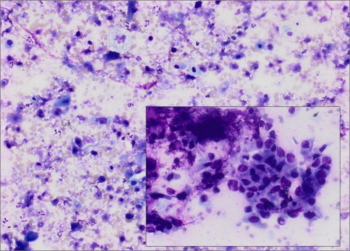
| Necrotic samples | Caution: Ensure that atypical single cells are not present within the necrotic debris,At ROSE necrotic slides can be misinterpreted as cellular. Review on high power to ensure the cells are viable. |
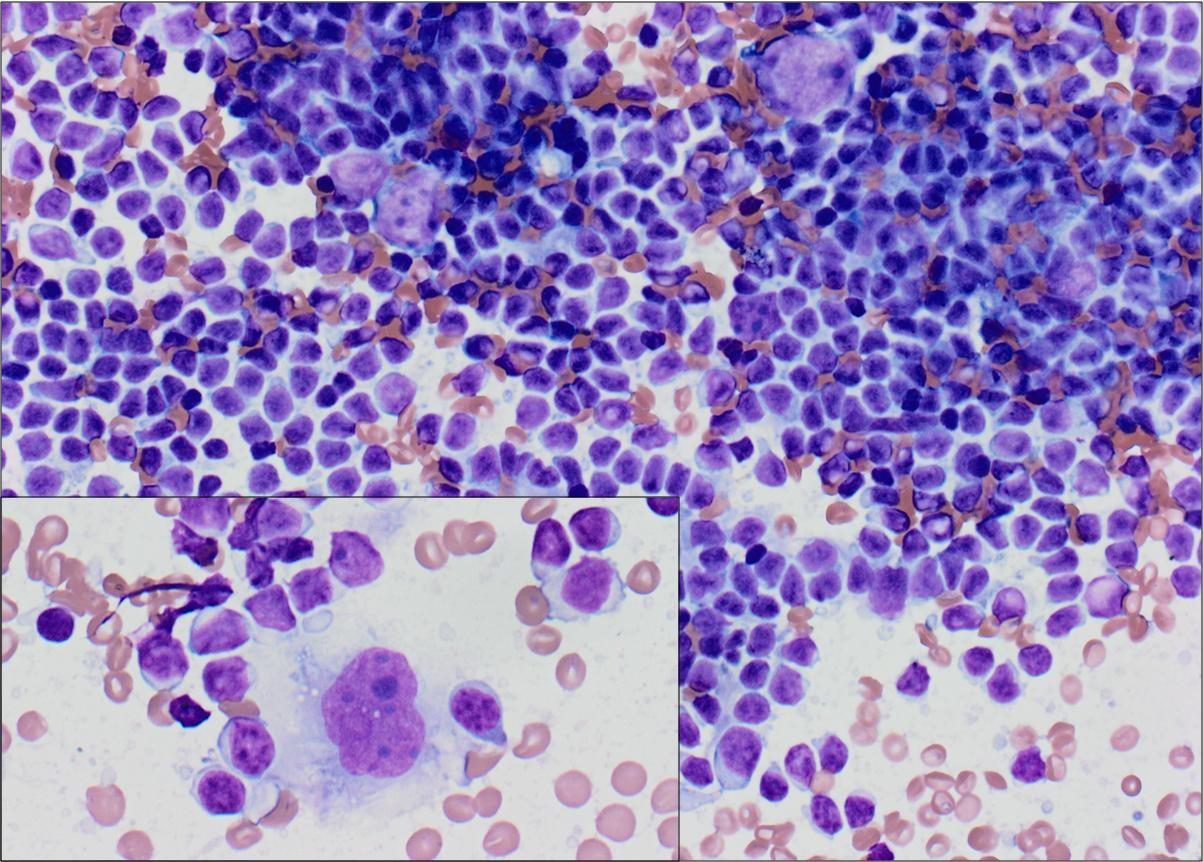
| Lymphoma cases | If possible, use 19/20 Gauge procore needle to ensure there are large cores for IHC panels – Ensure cores are filtered and separate from induced clot. Histology prepare the cores (worms) differently to prevent cutting through the section.If possible, use 19 Gauge procore needle to ensure there are large cores for IHC panels. An abundance of lymphocytes is not seen in benign node sampling is seen at ROSE. In some cases, highly atypical cells are also present |
| GI contaminant – epithelial cells and mucin | Must see both ductal and acinar cells present to confirm pancreatic sampling. Intestinal mucin is more metachromaticMust see both ductal and acinar cells present to confirm pancreatic sampling. Intestinal mucin is more metachromatic |
High power view is required in necrotic lesions. Do not assume all cells are necrotic
Adapted from: Glinski et al. Single slide assessment: A highly effective cytological rapid on?site evaluation technique for endobronchial and endoscopic ultrasound?guided fine needle aspiration. Cytopathology 30, 164–172 (2019).