Chronic lymphatic leukaemia/small lymphocytic lymphoma (CLL/SLL)
- Incidence: 7-8% of all NHL.
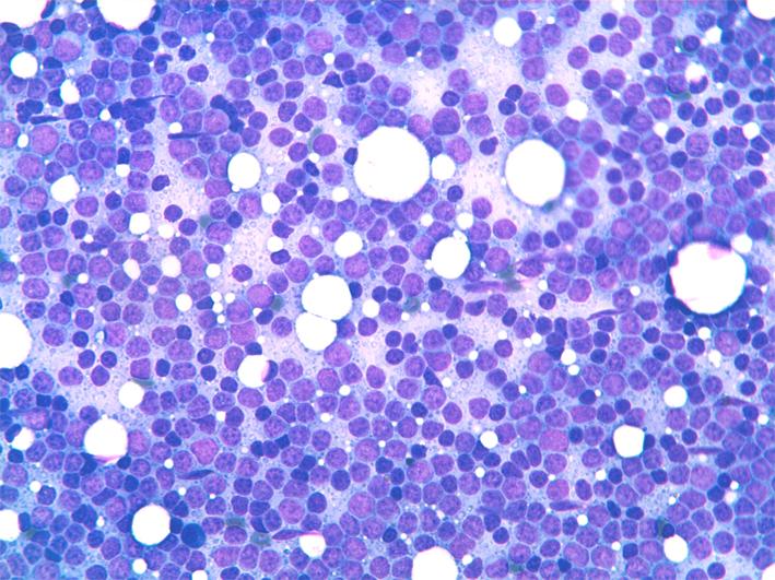
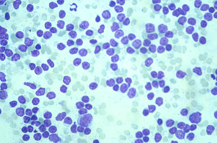
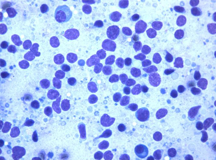
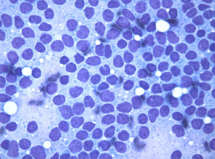
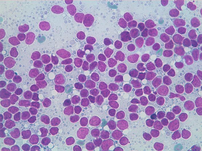
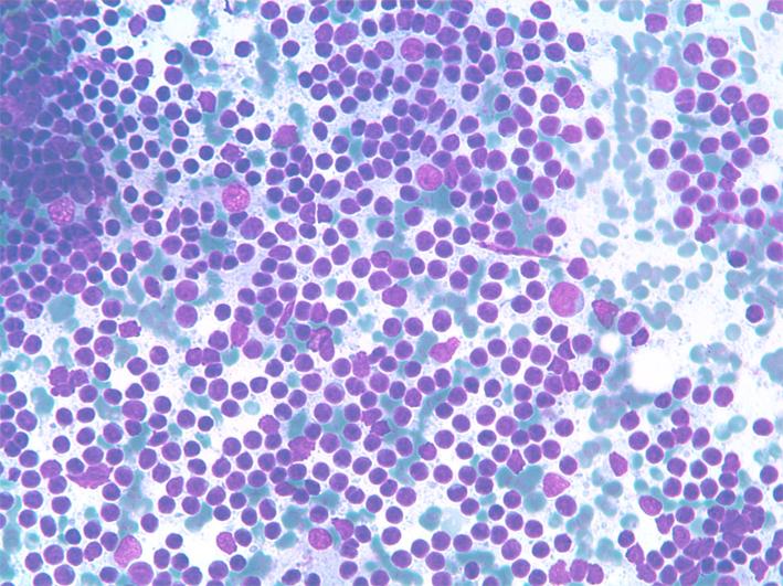
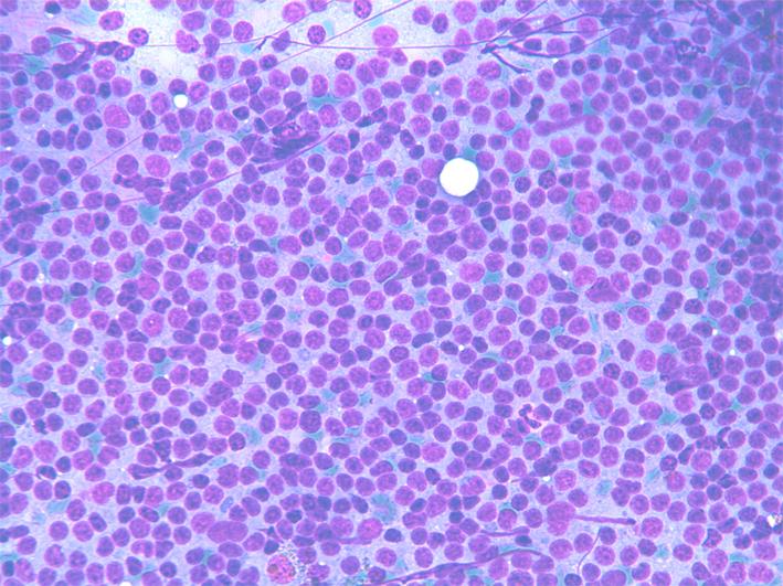
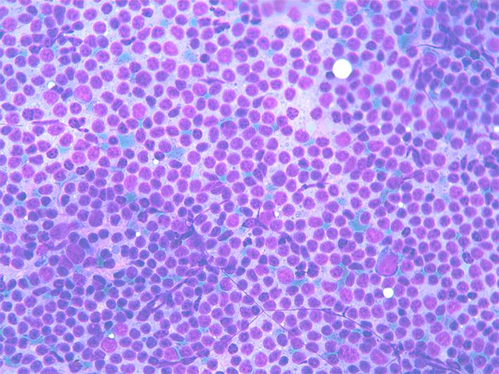
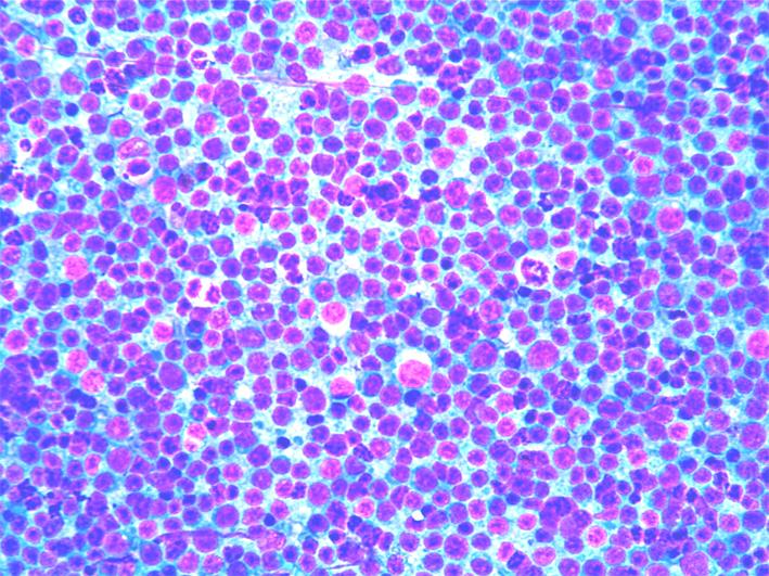
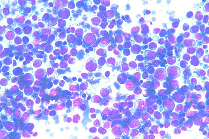
- Cytology: Smears show monotonous, dispersed cell populations composed of small lymphocytes with clumped chromatin and occasional, small, nucleoli; mitoses are absent or rare. Accelerate phases of CLL/SLL show larger, nucleolated, cells (paraimmunoblasts) and mitoses, and should not be confused with Richters syndrome, which is a large-cell histiocytic lymphoma arising on a CLL/SLL background.
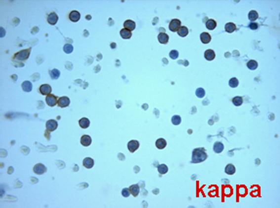
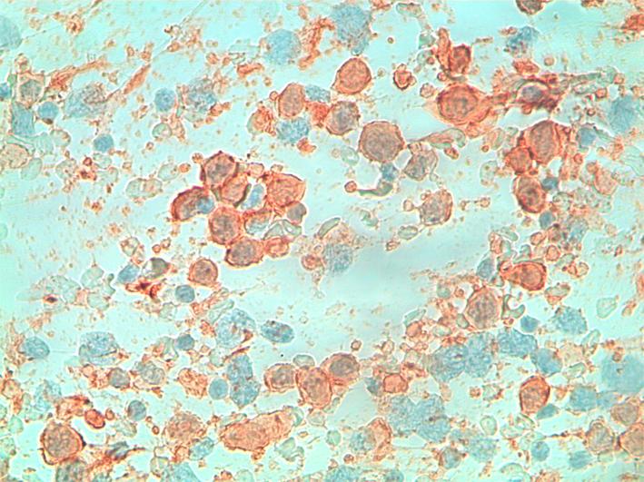
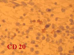
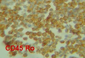
- ICC: light chain restriction, CD5+, CD20+, CD23+; Ki 67 is positive in a small percentage of the cells.
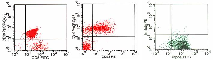
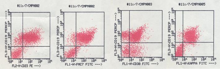
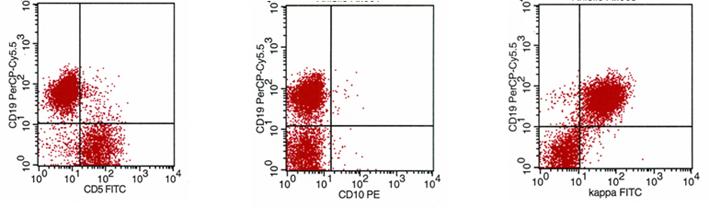
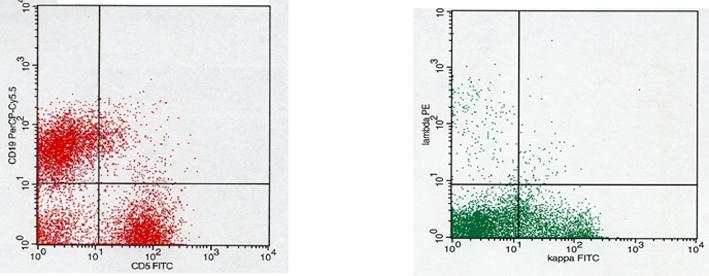
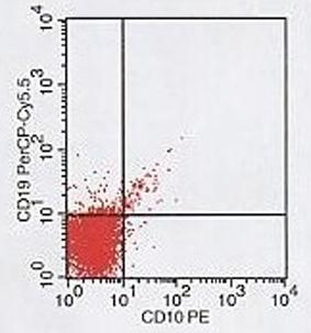
- FC: CD5/CD19+, CD19/CD23+, CD19+/CD10-, ? or ? light chain restriction.
- FISH: trisomy 12, 11q22 (ATM), 12, 13q14.3, 13q34.3 (LAMP1), and 17p13.1 (p53) as prognostic markers.
- Differential diagnosis: Non-specific hyperplasia ML, FL, MZL.
Lymphoplasmacytoid Lymphoma, Immunocytoma (LPCL)
- Incidence: 1-2% of all NHL.
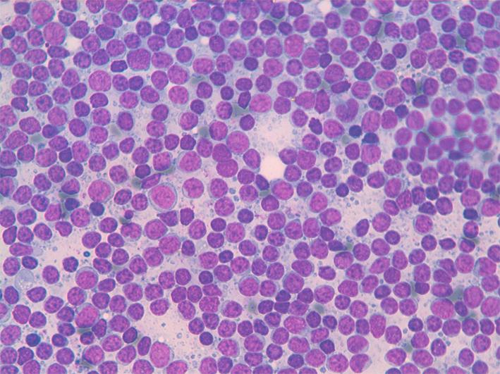
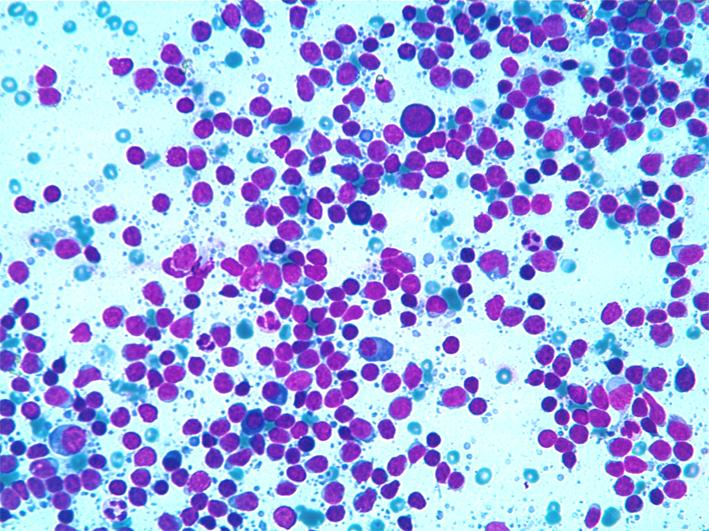
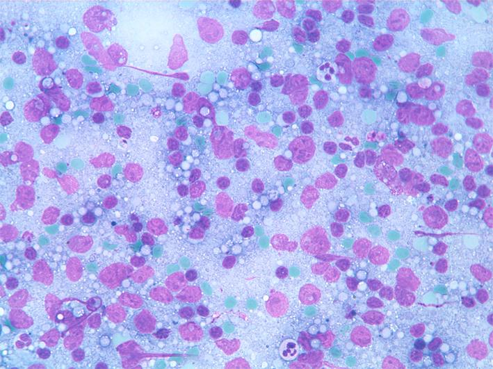
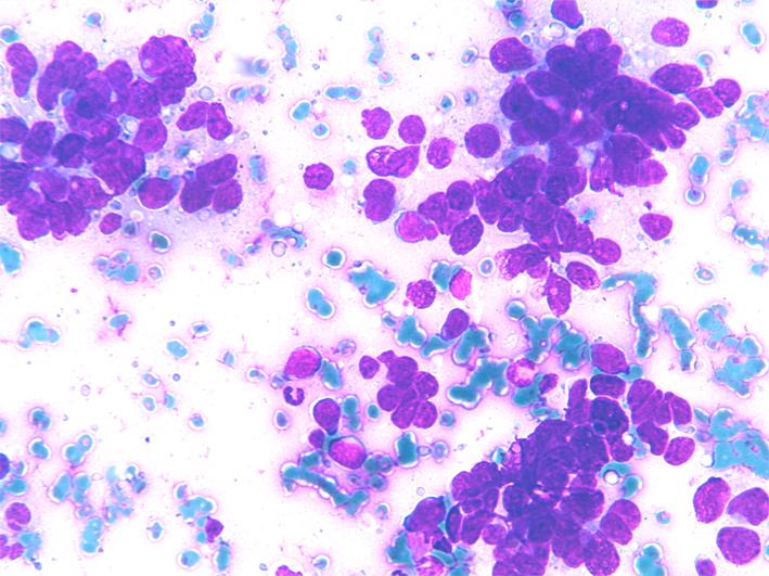
- Cytology: dispersed cell population composed of small lymphocytes with plasmacytoid differentiation; mature plasma cells are also present.
- ICC: CD20+, CD5-,CD10-, CD23-, Ki67+ in a variable percentage (10-20%) of cells.
- FC: CD5-/CD19+, CD19+/CD23+, CD19+/CD10-, CD19+/CD38+, ? or ? light chain restriction.
- Differential diagnosis: Non-specific hyperplasia, SLL/CLL.

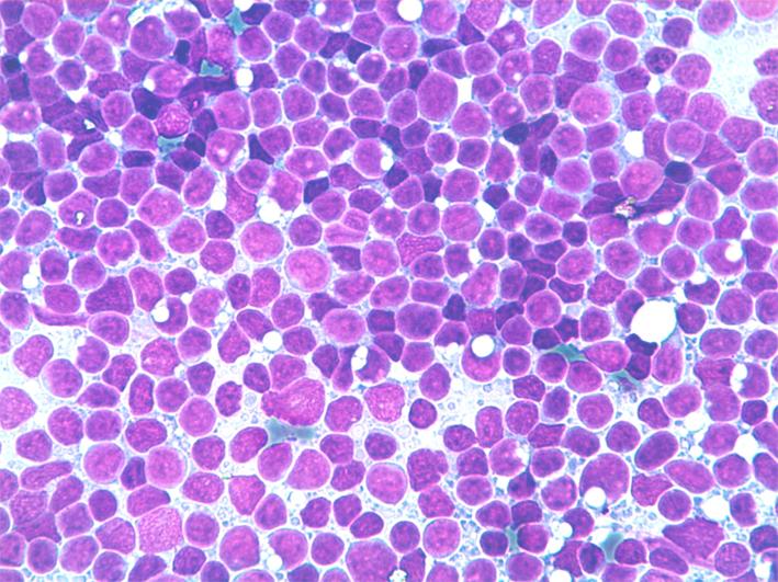
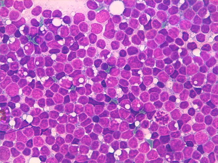
Mantle cell lymphoma (MCL)
- Incidence: 7-10% of all NHL.
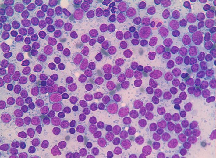
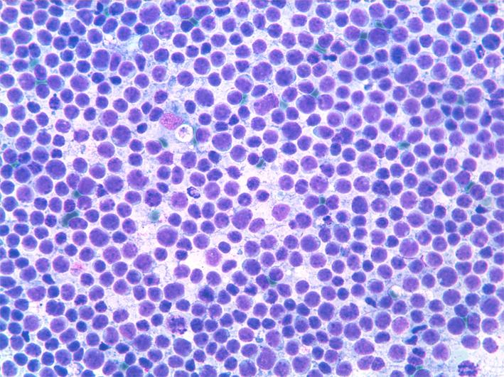
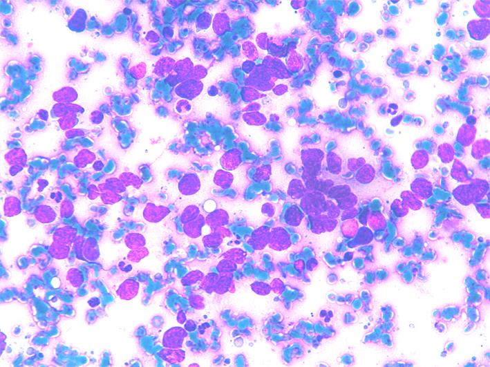
- Cytology: dispersed cell population composed of small/medium-size lymphocytes with irregular nuclei and a thin rim of pale cytoplasm. Nuclei may have slightly irregular shape, clumped chromatin and small nucleoli, or may be cleaved. Mitoses are scanty whereas they may be numerous in the blastoid variant
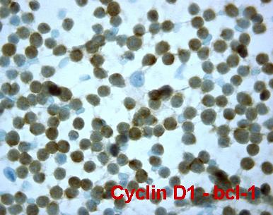
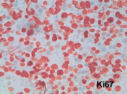
- ICC. CD20+, CD5+,CD10-, CD23-, Ki67+ in a variable percentage (10-20%) of the cells.
- FC: CD5+/CD19+, CD19/FMC7+, CD19+/CD23-, CD19+/CD10-, ? or ? light chain restriction.
- FISH: t(11;14)(q13;q32).
- Differential diagnosis: Non-specific hyperplasia, SLL/CLL,FL, MZL.

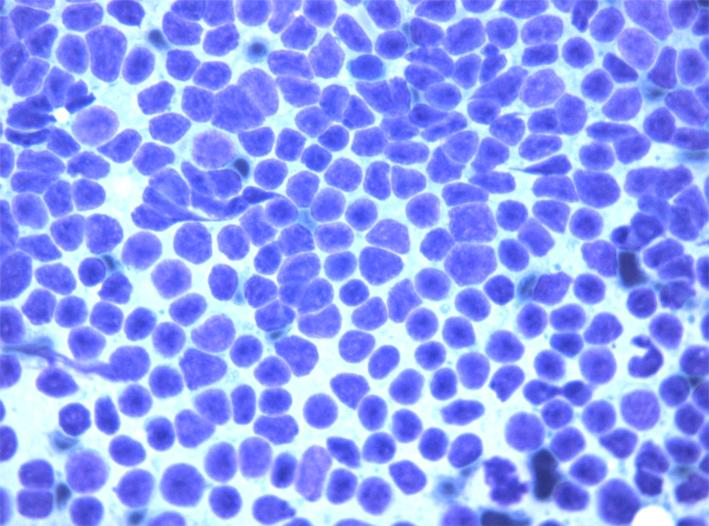
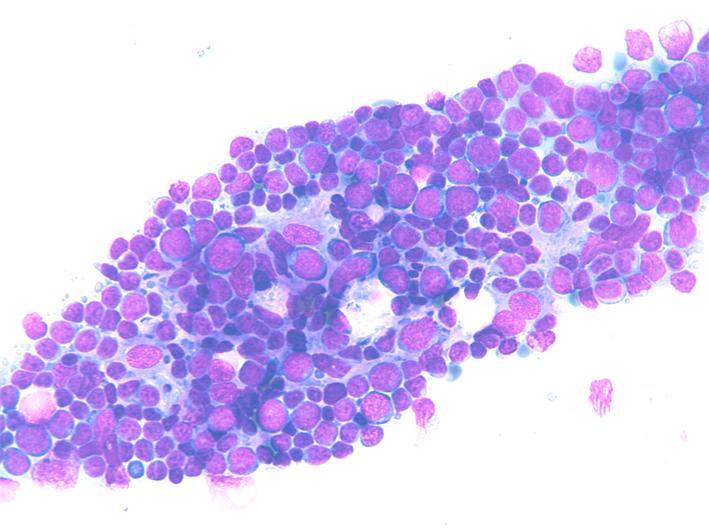
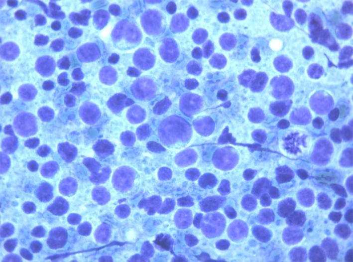
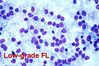
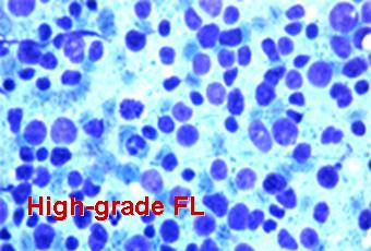
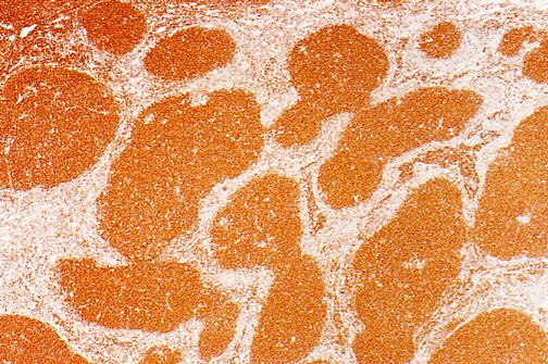
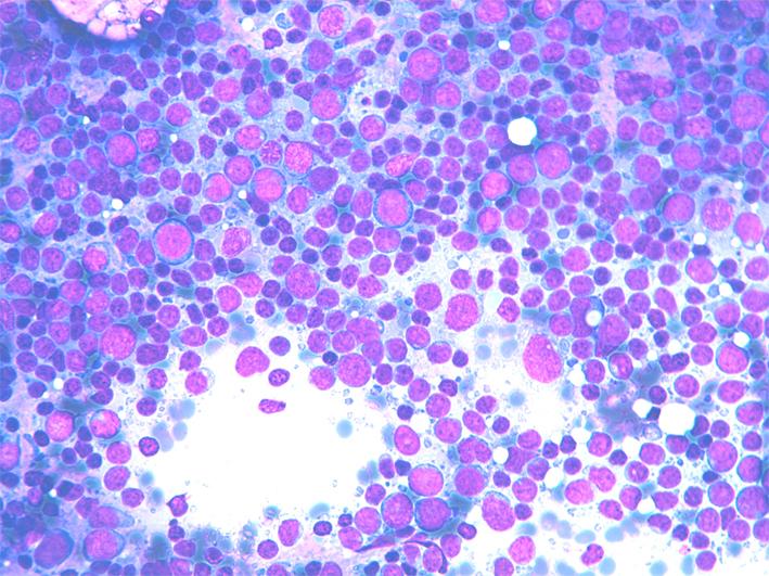
Follicular lymphoma (FL)
Follicular lymphoma (FL): cytology
- Incidence: 22-30% of all NHL.
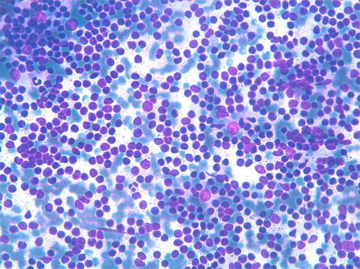
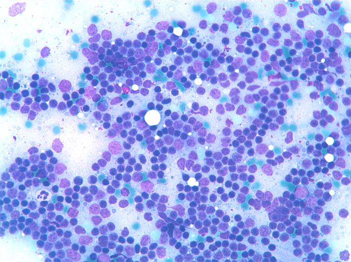
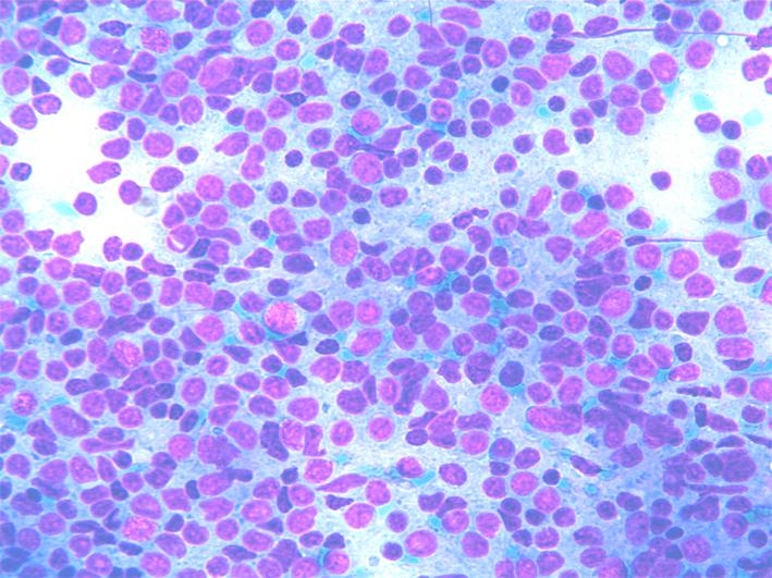
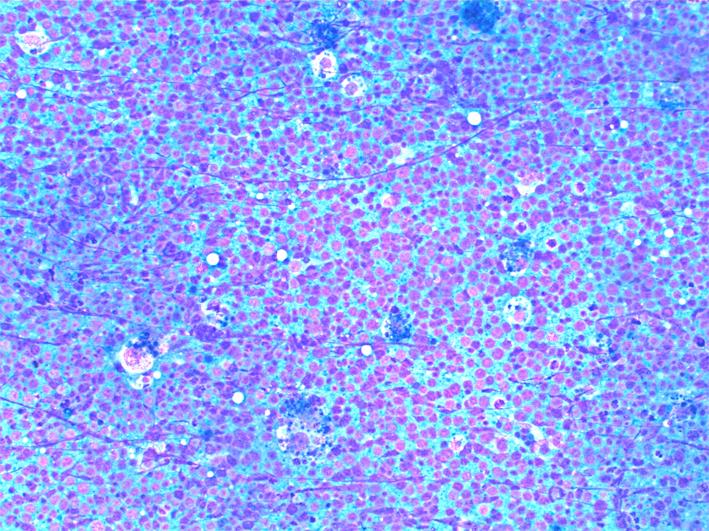
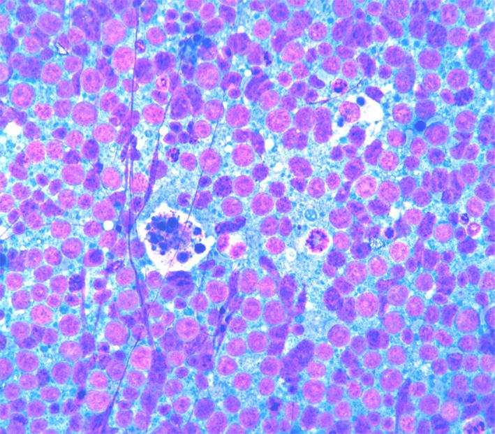
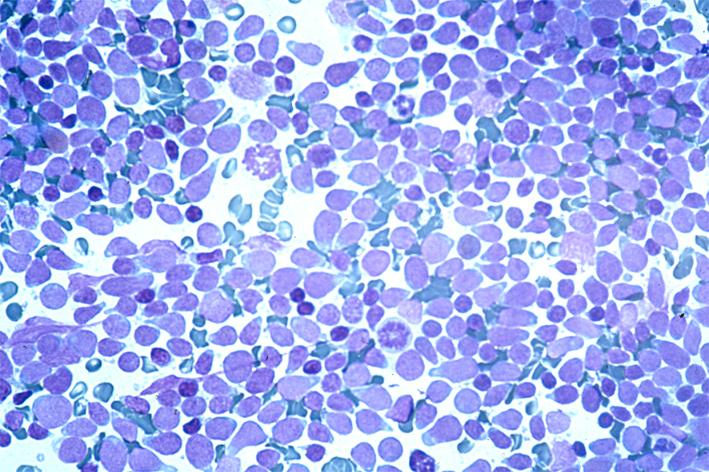
- Cytology: dispersed cell population composed of a spectrum of medium- to large-size lymphocytes, with irregular nuclei and a thin rim of pale cytoplasm. Nuclei may have slightly irregular shape, or be cleaved with clumped chromatin and inconspicuous nucleoli in grade I, to large nuclei with dispersed chromatin and two or more marginal nucleoli in grades II and III.
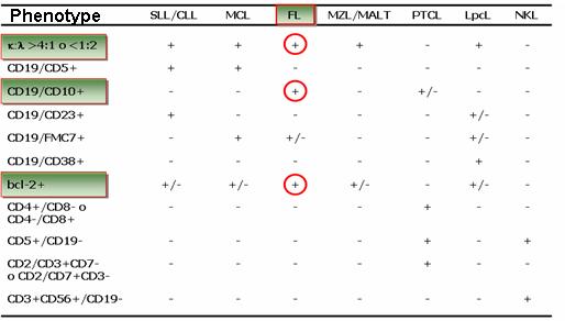
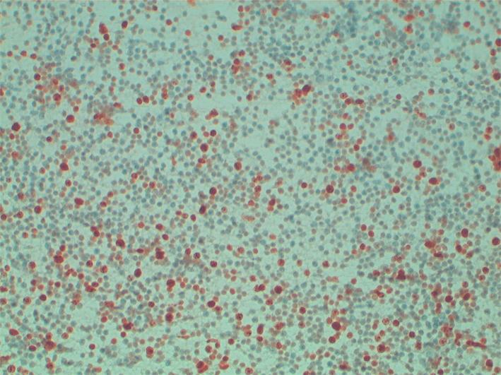
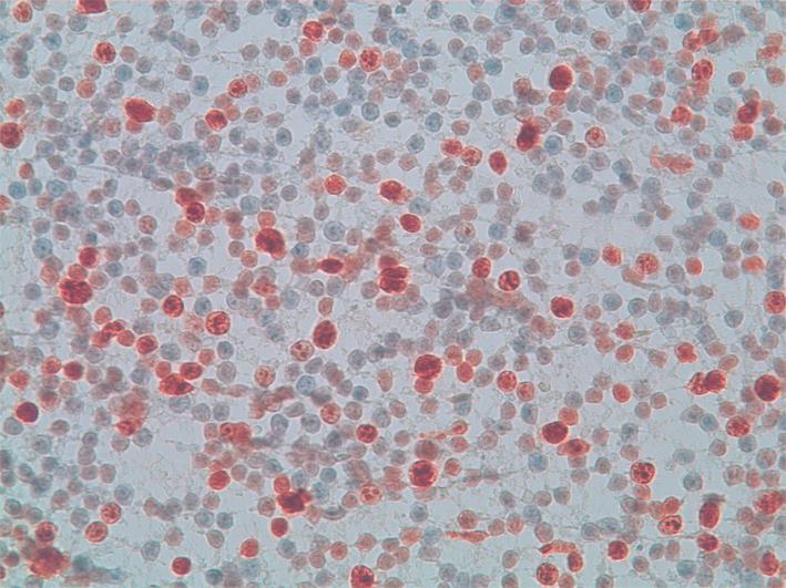
- ICC: CD20+,CD5-,CD10+,CD23-,Ki67+ in a variable percentage of the cells.
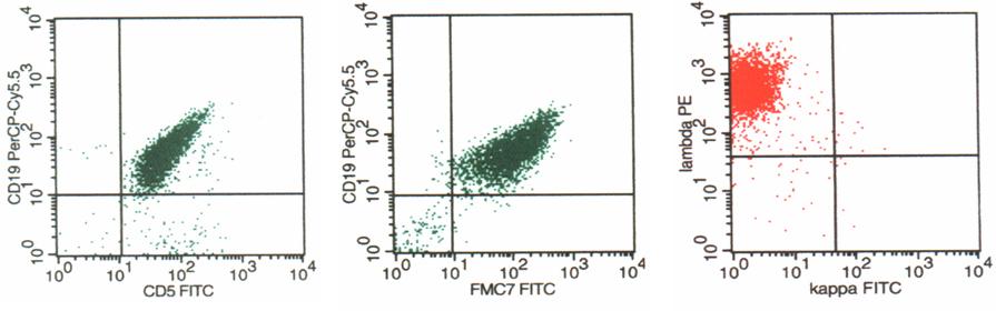
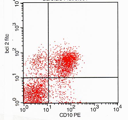
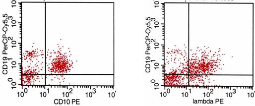
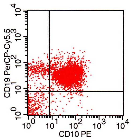
- FC: CD5-/CD19+, CD19+/FMC7+, CD19+/CD23-, CD19+/CD10+, CD10/bcl-2+, ? or ? light chain restriction.
- FISH: t(14;18)(q32;q21).
- Differential diagnosis: Non-specific hyperplasia, SLL/CLL,MCL.

Follicular lymphoma (FL): flow cytometry
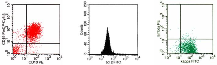
and one or more small nucleoli. FC shows CD10/bcl-2 co-expression that reflects the follicles bcl-2 histological positivity.
and one or more small nucleoli. FC shows CD10/bcl-2 co-expression that reflects the follicles bcl-2 histological positivity.
and one or more small nucleoli. FC shows CD10/bcl-2 co-expression that reflects the follicles bcl-2 histological positivity.
and one or more small nucleoli. FC shows CD10/bcl-2 co-expression that reflects the follicles bcl-2 histological positivity.
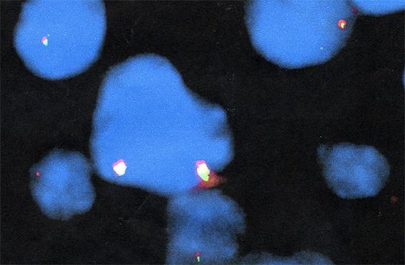
Follicular lymphoma (FL): FISH
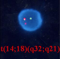
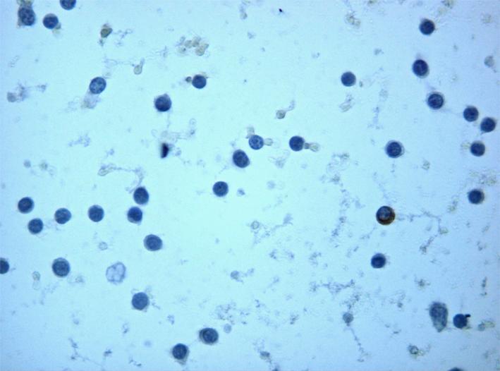
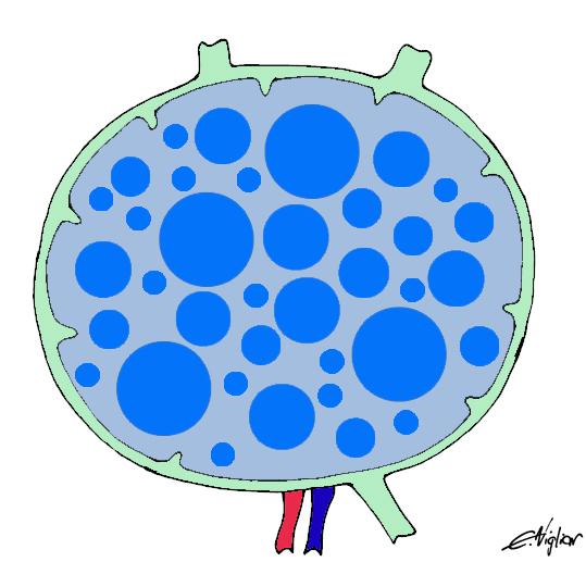
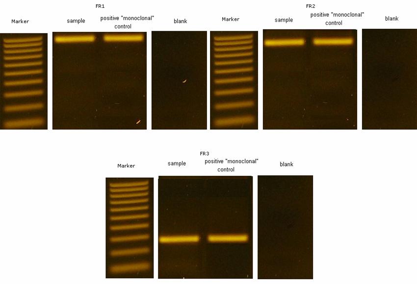
In cases of FL (CD10/19 co-expression) with dim or no light chains expression, FISH may be helpful in diagnosis by the demonstration of t14-18. Bcl2 locus (18q21,labelled red), overlaps IGH locus (14q32, labelled in green) giving a signal of fusion (yellow).
IGH gene rearrangement in lymphoma with dim or no light chains expression
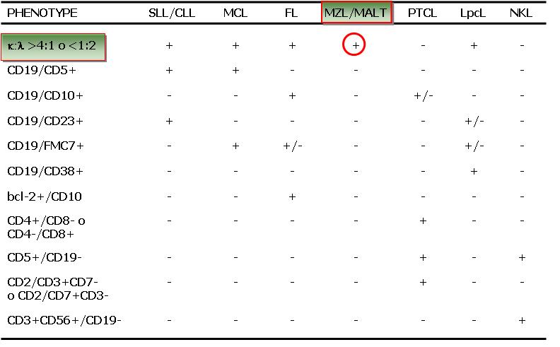
Marginal Zone B-cell Lymphoma (MZL)
- Incidence: 2-3% of all NHL.
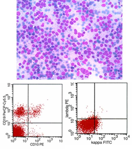
- Cytology: dispersed cell population composed of small lymphocytes with irregular nuclei. Nuclei may have slightly irregular shape, dense chromatin and indistinct nucleoli, or be cleaved. The rim of pale cytoplasm observed in histological sections is rare or undetectable on the smears. Monocytoid cells and plasma cells may be intermingled, mitoses are scanty.
- ICC: CD20+, CD5-,CD10-, CD23-, Ki67+/-.
- FC: CD5-/CD19+, CD19+/FMC7-, CD19+/CD23-, CD19+/CD10-, ? or ? light chain restriction.
- Differential diagnosis: Non-specific hyperplasia, SLL/CLL,MCL, FL.
Extranodal B-cell lymphoma of mucosa associated lymphoid tissue (MALT)
Diffuse large B-cell lymphoma (DLBCL)
- Incidence: 30-40% of all NHL.
- Cytology: dispersed cell population composed of large lymphoid cells with irregular nuclei, one or more large nucleoli and a wider rim of pale cytoplasm. Morphological variants of DLBCL are centroblastic, immunoblastic T-cell/histiocyte-rich and anaplastic. Nuclei may be slightly irregular in shape or cleaved and have dense chromatin and indistinct nucleoli. Lymphocyte and plasma cells may be intermingled, Mitoses are scanty.
- ICC: CD19+, CD20+, 22+, CD79a+, Ki67+ in a variable percentage (10-20%) of the cells.
- FC: CD5-/CD19+, CD19+/FMC7-, CD19+/CD23-, CD10-, ? or ? light chains restriction.
- DD: HL, metastases.

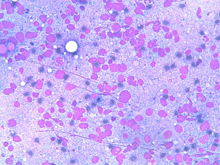
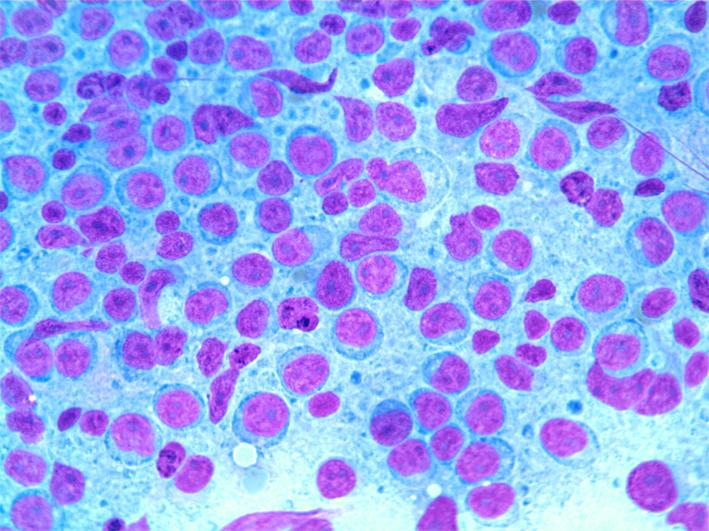
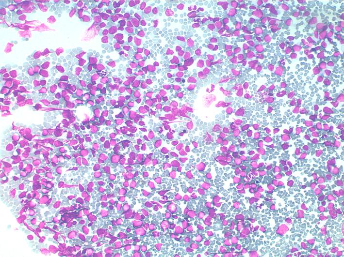
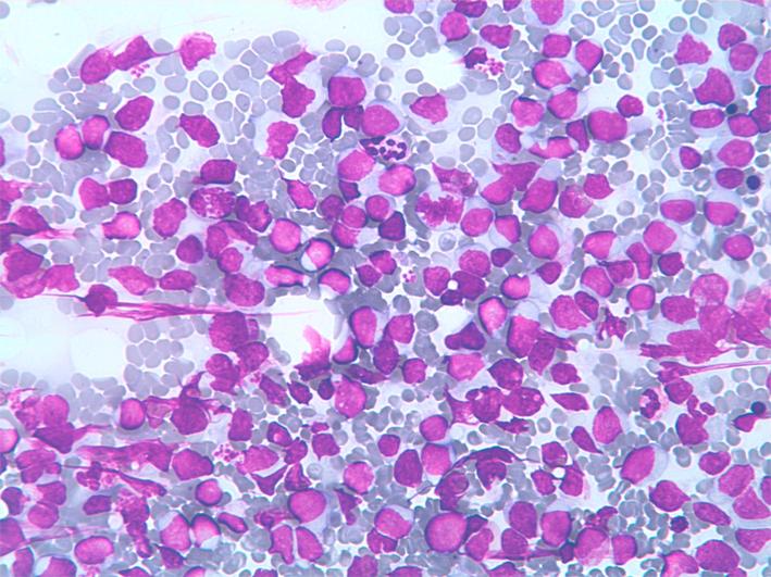
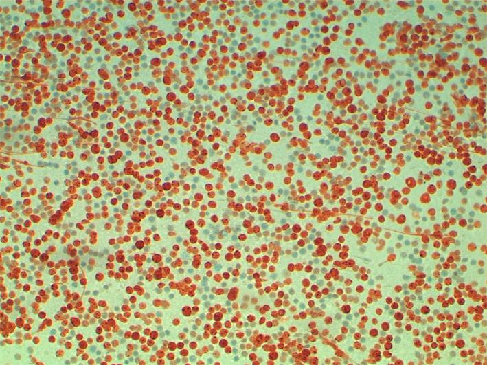
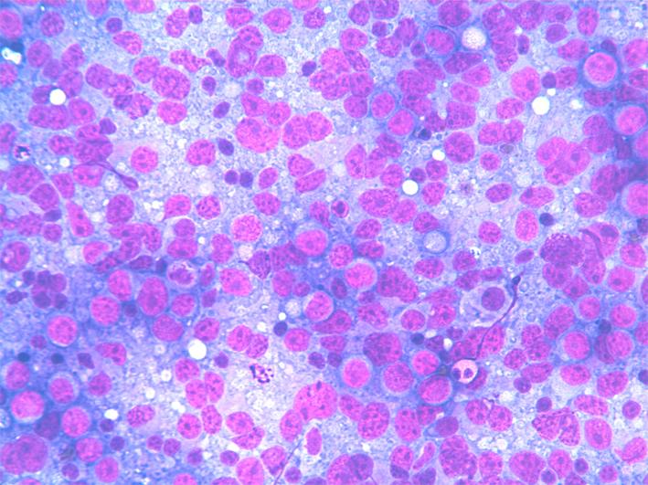
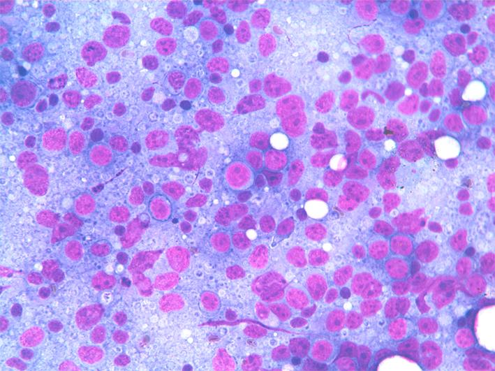
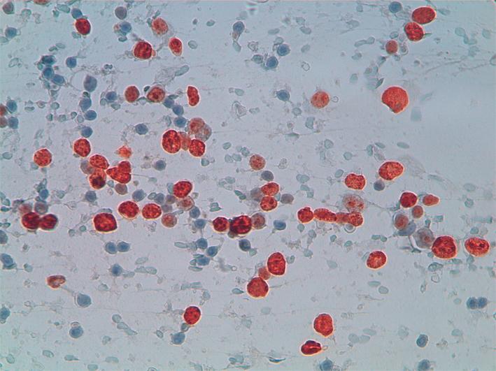
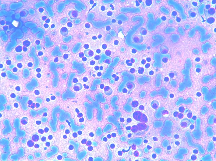
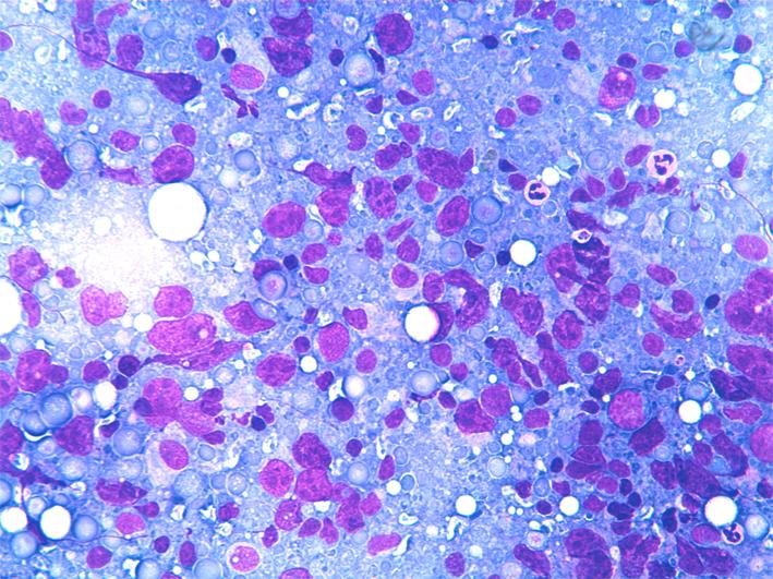
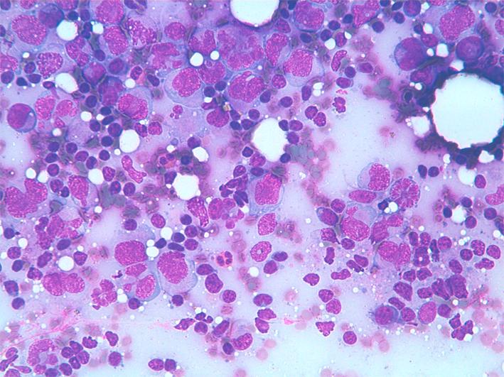
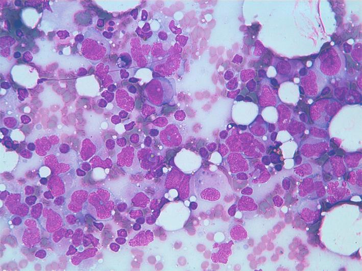
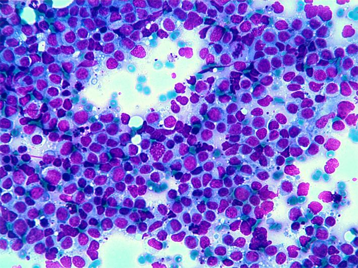
Plasma cell myeloma/plasmacytoma
- Incidence: 15% of haematological malignancies.
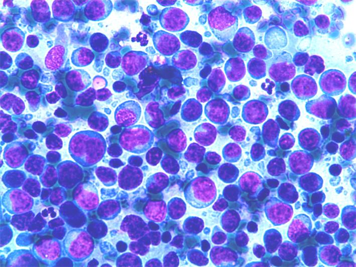
- Cytology: in typical cases show dispersed cell population composed of small/medium-size plasma cells. Cells show one or two nuclei with clumped cartwheel chromatin and small nucleoli. Cytoplasm, when present are well defined with evident perinuclear Golgi apparatus. In poorly differentiated cases nuclei are atypical and may show marked anisonucleosis.
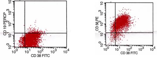
- ICC. CD20-/+, CD5-, CD10-, CD23-, CD38, CD79a, EMA, Ki67+ in a variable percentage.
- FC: CD38+, CD38/56 +/-, CD10-, ? or ? light chain restriction.
- Differential diagnosis: non-haematological tumours in poorly differentiated cases.


Burkitt lymphoma (BL)
- Incidence: 1-2% of all NHL.
- Cytology: dispersed cell population composed of undifferentiated medium sized lymphocytes with dispersed chromatin and one or more small basophilic nucleoli; the cytoplasm is basophilic and sometimes vacuolated. Immature nuclei are fragile, resulting in frequent nuclear crushes in the smear; a high mitotic index is observed. Scattered macrophages with engulfed cytoplasm may give a starry sky pattern to the smear.
- ICC: CD20+, CD5-, CD10+, Bcl-6+, CD23-, Ki67+ in a high percentage of the cells.
- FC: CD5-/CD19+, CD19+/FMC7-, CD19+/CD23-, CD19+/CD10+, light chain often unexpressed.
- FISH: t(8;14)(8q24;q32).
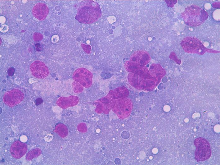
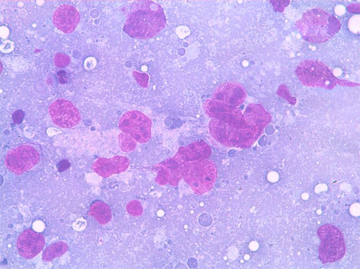
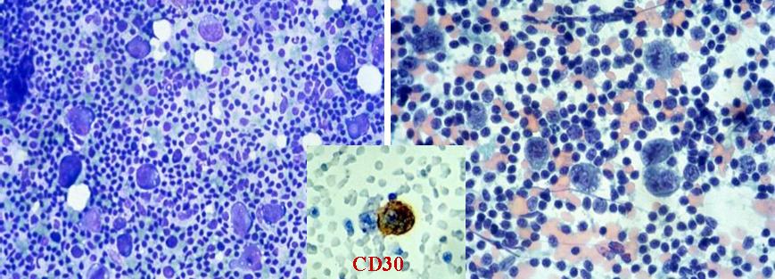
Anaplastic large cell lymphoma
- Incidence: 3% of all NHL.
- Cytology: dispersed and polymorphous large cells with very irregular shape. Multilobated, horseshoe bi- and multinuclecleated cells are present. The nuclei have coarse chromatin and larger nucleoli.
- Background is generally polymorphous with mature lymphocytes, granulocytes and eosinophils. Atypical mitoses and necrosis may be seen.
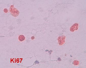
- ICC: CD20-, CD45+ CD15-, CD30++, Ki67+, EMA+, ALK-1+/-.
- FC: CD5+/-, CD19+/-, aberrant phenotypes may be observed.
- FISH: t(2;5)(p23;q35).
- PCR: clonal rearrangement of the T-cell receptor (90%).
- Differential diagnosis: DLBCL, HL, metastases.
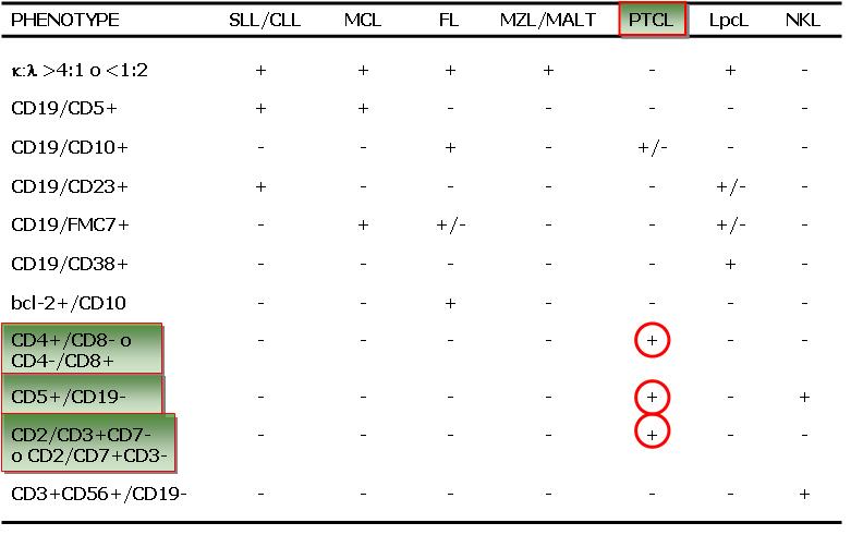
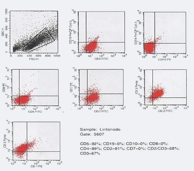
Peripheral T-cell lymphoma (PTL)
- Incidence: 7-8% of all the NHL.
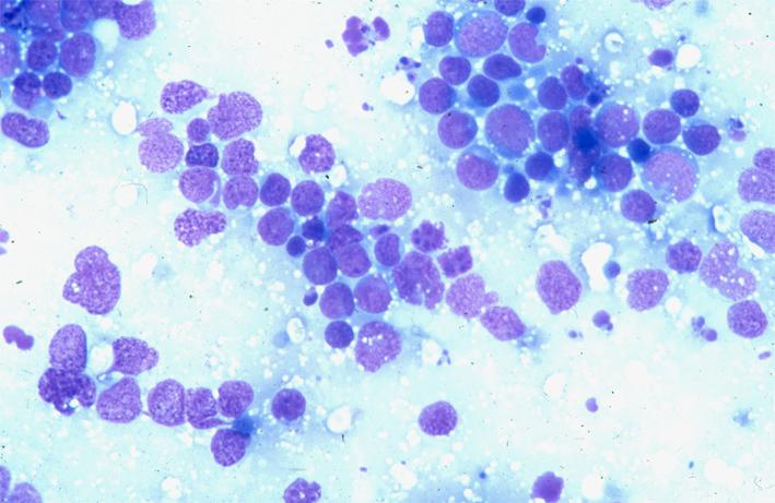
- Cytology: dispersed and polymorphous medium- or large-sized cells with irregular shape, vesicular nuclei and evident nucleoli. The nuclei have irregular shape, coarse chromatin and one or more large nucleoli. Binucleated cells are frequently observed. The background is generally polymorphous; mature lymphocytes, granulocytes, numerous eosinophils may be present.
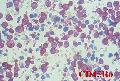
- ICC: CD20-, CD3+, CD4+ CD10-, CD23-, Ki67+ in a variable percentage of the cells.
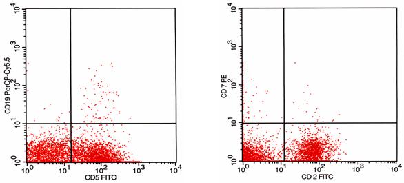
- FC: CD5+, CD19-, CD10-, CD2+ CD3+ CD7-.
- PCR: TC receptor rearranged in most cases.
- Differential diagnosis: B-cell NHL.

Precursor T-lymphoblastic lymphoma/leukemia (TLL)
- Incidence: <1% of all the NHL.
- Cytology: dispersed and polymprphous medium-sized undifferentiated cells. Nuclei are round or convoluted with dispersed chromathin and small nucleoli. High mitotic index and nuclear fragility.
- ICC: CD20-, CD3+, CD10-, TDT+, Ki67+ in a high percentage of the cells.
- FC: CD5 +/-, CD19-, CD10-, CD2+ CD3+ CD7-.
- Differential diagnosis: BL.
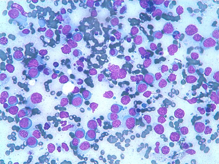
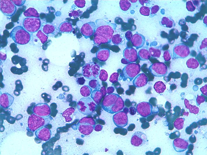
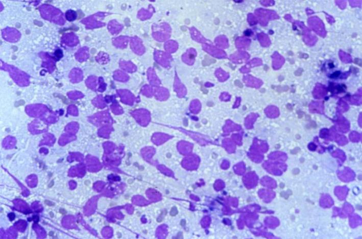
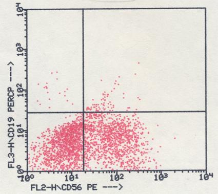
NK-cell lymphoma/leukemia
- Incidence: <1% of all the NHL.
- Cytology: dispersed and monomorphous small- or medium-sized undifferentiated cells. The nuclei are round or convoluted with dispersed chromatin and small nucleoli. There is also a high mitotic index and nuclear fragility.
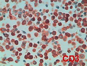
- ICC: CD20-, CD3+, CD10-, TDT+, CD56+ Ki67+ in a high percentage of the cells.
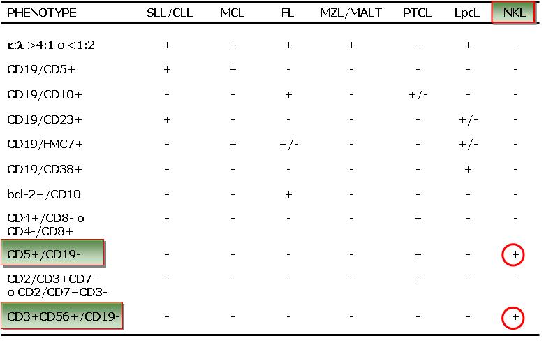
- FC: CD56+, CD5 -, CD19-, CD10-, CD2-CD3- CD7-.
- Differential diagnosis: PTL, LLA, TLL.

Blastic NK-cell lymphoma/leukemia