Uno striscio cervicale che è ottenuto utilizzando una spatola o comunque uno strumento in modo corretto contiene una ampia quantità di cellule normali intrappolare nel muco cervicale. Le cellule epiteliali possono essere normalmente esfoliate o ottenute da un’energico passaggio della spatola sulla regione dell’ostio esterno.
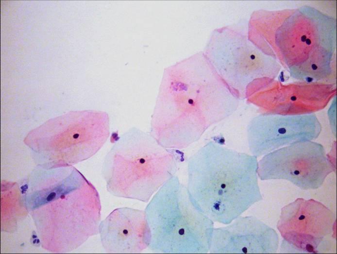
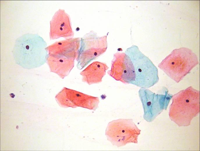
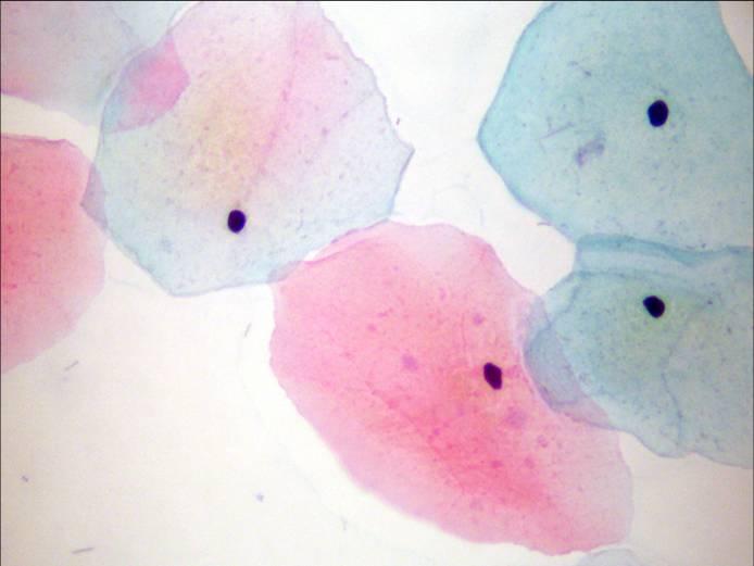
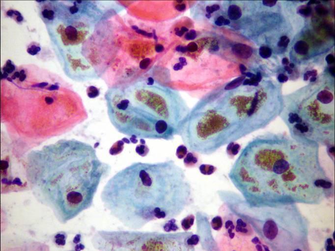
SUPERFICIAL SQUAMOUS CELLS: These cells are derived from mature squamous epithelium which has developed to full thickness under the influence of oestrogen. They appear as large polygonal squames with transparent cytoplasm and angular borders. Folding is rare. They measure 45-50 u in diameter. The nuclei are small condensed and pyknotic with no visible nuclear structure and do not exceed 5u. Karyorrhexis is sometimes seen. The cytoplasm stains pink or orange with occasional effete cells staining blue.SUPERFICIAL CELLS: Note orangophillic stain with effete cells staining blue. Compare the size of the squames with the size of the polymorphs (5-10u). Superficial squames are usually found mid cycle.SUPERFICIAL CELLS: note keratohyaline graules in cytoplasm. Keratinisation is not a feature of a normal cervix. Single cells reflect spontaneous exfoliation of surface epithelium Sheets of superficial squames reflect detachment of surface layer with smear taking device.INTERMEDIATE SQUAMOUS CELLS: These are shed from semi mature squamous epithelium and reflect the effect of progesterone (or oral contraceptive) or diminished oestrogen. These flat cells are slightly smaller than the superficial squamous cells (35- 40u) and are less angular. The nuclei are vesicular and 8u diameter and have a delicate chromatin network. The cytoplasm is cyanophilic and denser than that of the superficial cells.INTERMEDIATE CELLS; sheets of overlapping intermediate cells which have been forcibly detached by the spatula. Note vesicular nuclei and lacto bacilli (doderlein bacilli) over lying the cells.INTERMEDIATE CELLS. In the late secretory phase of the menstrual cycle and in pregnancy the morphology of the intermediate cells alters. They appear boat shaped, with thickened cytoplasmic borders and eccentric nuclei. The cells are sometimes described as navicular cells and contain abundant glycogen which appears brownish in the light microscope.PARABASAL CELLS: Parabasal cells are the predominant cell type in atrophic or post partum smears. They are small (15-30u) with dense cyanophilic cytoplasm. Effete eosinophilic cytoplasmic staining is occasionally seen Nuclei have a fine chromatin pattern and occupy about one half of the cell. The cells are spontaneously exfoliated they appear rounded. When they have been forcibly detached they appear as sheets of cells. Parabasal cells are particularly fragile and their cytoplasm may disintegrate during smear preparation. Basal cells are rarely seen in cervical smears because of their deep position in the mucosa and their attachment to the basement membrane.PARABASAL CELLS Sheets of parabasal cells in an atrophic smear. Note occasional bare nuclei and mucous strands.
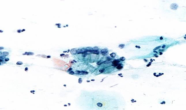
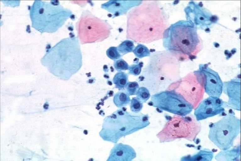
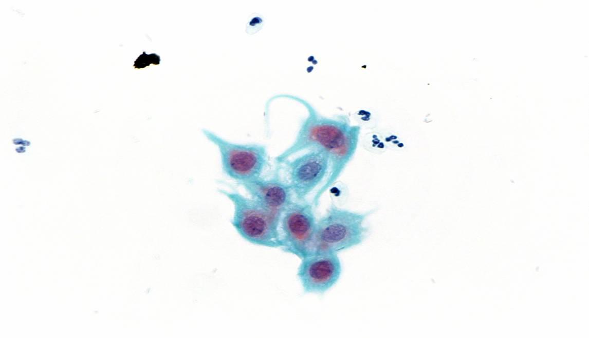
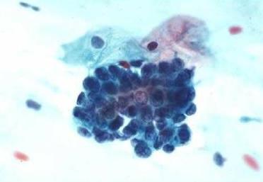
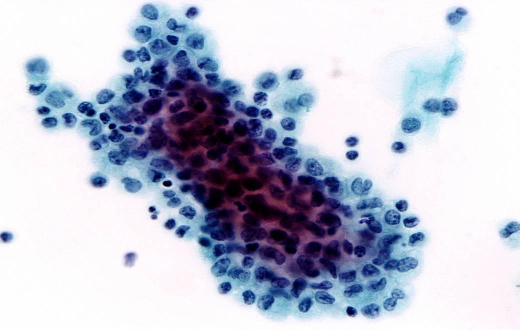
Cellule derivate dall’epitelio colonnare del canale endocervicale
ENDOCERVICAL GLANDULAR EPITHELIAL CELLS: These are columnar shaped cells with basal nuclei and vaculotated delicate cytoplasm. The chromatin is fine and one or more nucleoli may be present. They may appear as strips (in palisade) or as a honey comb pattern. They are very fragile and may be poorly preserved even in a well taken smear. Naked nuclei may be seen. Discrete endocervical cells seen “end on” can be distinguished from discrete parabasal cells by their eccentric nuclei and delicate cytoplasm.ENDOCERVICAL GLANDULAR EPITHELIAL CELLS: These are columnar shaped cells with basal nuclei and vaculotated delicate cytoplasm. The chromatin is fine and one or more nucleoli may be present. They may appear as strips (in palisade) or as a honey comb pattern. They are very fragile and may be poorly preserved even in a well taken smear. Naked nuclei may be seen. Discrete endocervical cells seen “end on” can be distinguished from discrete parabasal cells by their eccentric nuclei and delicate cytoplasm.ENDOCERVICAL GLANDULAR EPITHELIAL CELLS: These are columnar shaped cells with basal nuclei and vaculotated delicate cytoplasm. The chromatin is fine and one or more nucleoli may be present. They may appear as strips (in palisade) or as a honey comb pattern. They are very fragile and may be poorly preserved even in a well taken smear. Naked nuclei may be seen. Discrete endocervical cells seen “end on” can be distinguished from discrete parabasal cells by their eccentric nuclei and delicate cytoplasm.ENDOCERVICAL GLANDULAR EPITHELIAL CELLS: These are columnar shaped cells with basal nuclei and vaculotated delicate cytoplasm. The chromatin is fine and one or more nucleoli may be present. They may appear as strips (in palisade) or as a honey comb pattern. They are very fragile and may be poorly preserved even in a well taken smear. Naked nuclei may be seen. Discrete endocervical cells seen “end on” can be distinguished from discrete parabasal cells by their eccentric nuclei and delicate cytoplasm.
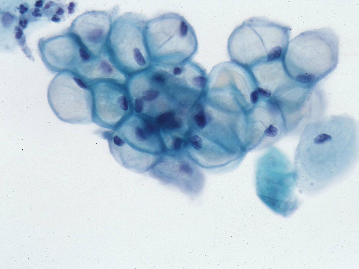
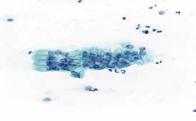
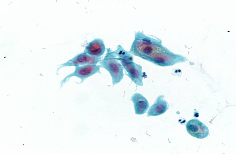
Cellule derivate dall’epitelio metaplastico della zona di trasformazione
IMMATURE SQUAMOUS METAPLASTIC CELLS: These cells are the size of parabasal cells (15-30u) and closely resemble them. The nuclei have a fine chromatin pattern and the cytoplasm is dense and cyanophilic; the n/c ratio is high. Immature metaplastic cells can be distinguished from atrophic parabasal cells by the fact that they are always found in association with mature superficial and intermediate squamous cells. NOTE: Mature metaplastic squamous epithelial cells cannot be distinguished from original (native) squamous epithelial cells in a cervical smear.IMMATURE SQUAMOUS METAPLASTIC CELLS: These cells are the size of parabasal cells (15-30u) and closely resemble them. The nuclei have a fine chromatin pattern and the cytoplasm is dense and cyanophilic; the n/c ratio is high. Immature metaplastic cells can be distinguished from atrophic parabasal cells by the fact that they are always found in association with mature superficial and intermediate squamous cells. NOTE: Mature metaplastic squamous epithelial cells cannot be distinguished from original (native) squamous epithelial cells in a cervical smear.IMMATURE SQUAMOUS METAPLASTIC CELLS: These cells are the size of parabasal cells (15-30u) and closely resemble them. The nuclei have a fine chromatin pattern and the cytoplasm is dense and cyanophilic; the n/c ratio is high. Immature metaplastic cells can be distinguished from atrophic parabasal cells by the fact that they are always found in association with mature superficial and intermediate squamous cells. NOTE: Mature metaplastic squamous epithelial cells cannot be distinguished from original (native) squamous epithelial cells in a cervical smear.IMMATURE SQUAMOUS METAPLASTIC CELLS: These cells are the size of parabasal cells (15-30u) and closely resemble them. The nuclei have a fine chromatin pattern and the cytoplasm is dense and cyanophilic; the n/c ratio is high. Immature metaplastic cells can be distinguished from atrophic parabasal cells by the fact that they are always found in association with mature superficial and intermediate squamous cells. NOTE: Mature metaplastic squamous epithelial cells cannot be distinguished from original (native) squamous epithelial cells in a cervical smear.
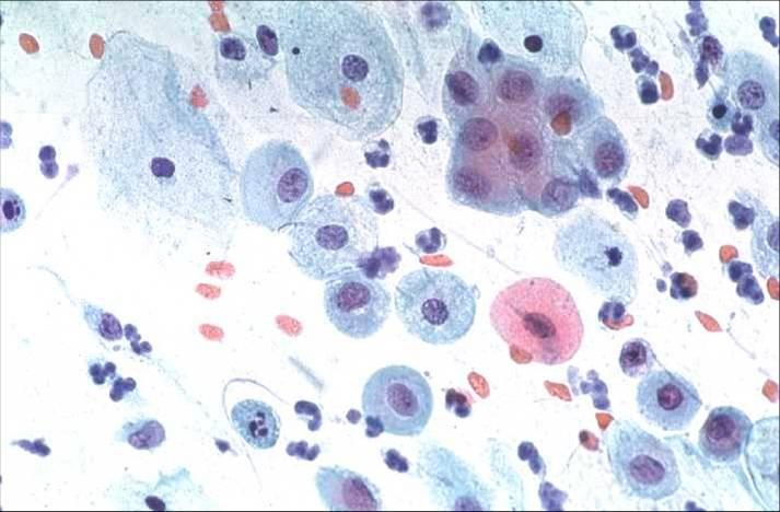
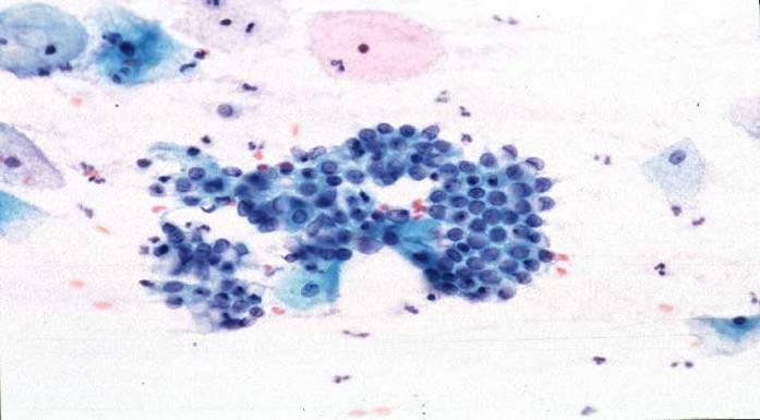
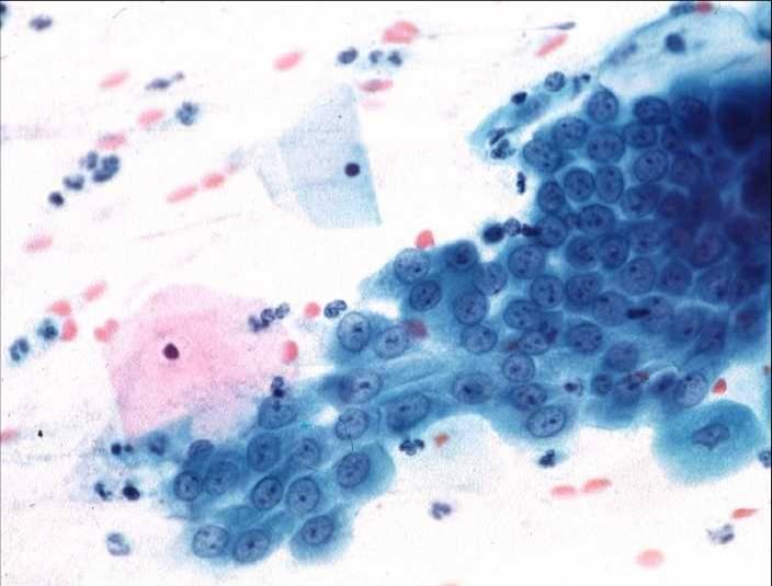
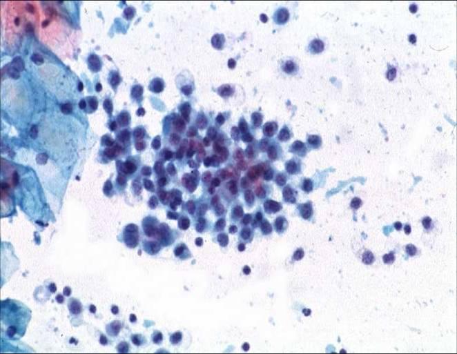
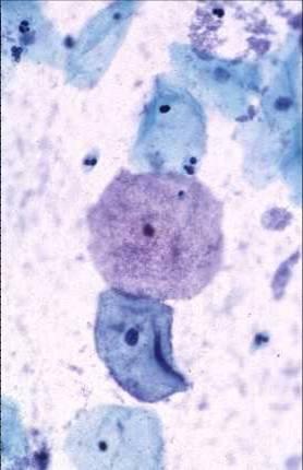
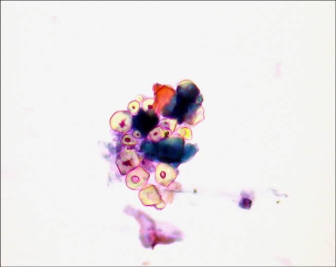
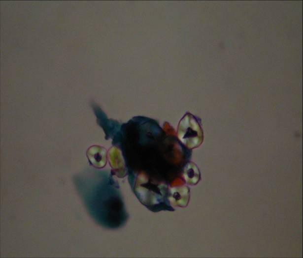
cellule endometriali
ENDOMETRIAL CELLS: Endometrial cells are a normal finding in smear taken during mensturation or in the first ten days of the menstrual cycle. Their presence at other times may have a pathological significance. They can be distinguished from endocervical cells by their small size (5-20u), cuboidal shape and rounded nuclei which often have a coarse chromatin structure and may vary slightly in size. They are often found in conjunction with histiocytes. They may present as single cells, or as rounded berry like structures. Alternatively they appear in menstural smears as a dense core of stromal cells surrounded by a paler fringe of glandular cells.ENDOMETRIAL CELLS: Endometrial cells are a normal finding in smear taken during mensturation or in the first ten days of the menstrual cycle. Their presence at other times may have a pathological significance. They can be distinguished from endocervical cells by their small size (5-20u), cuboidal shape and rounded nuclei which often have a coarse chromatin structure and may vary slightly in size. They are often found in conjunction with histiocytes. They may present as single cells, or as rounded berry like structures. Alternatively they appear in menstural smears as a dense core of stromal cells surrounded by a paler fringe of glandular cells.ENDOMETRIAL CELLS: Endometrial cells are a normal finding in smear taken during mensturation or in the first ten days of the menstrual cycle. Their presence at other times may have a pathological significance. They can be distinguished from endocervical cells by their small size (5-20u), cuboidal shape and rounded nuclei which often have a coarse chromatin structure and may vary slightly in size. They are often found in conjunction with histiocytes. They may present as single cells, or as rounded berry like structures. Alternatively they appear in menstural smears as a dense core of stromal cells surrounded by a paler fringe of glandular cells.ENDOMETRIAL CELLS: Endometrial cells are a normal finding in smear taken during mensturation or in the first ten days of the menstrual cycle. Their presence at other times may have a pathological significance. They can be distinguished from endocervical cells by their small size (5-20u), cuboidal shape and rounded nuclei which often have a coarse chromatin structure and may vary slightly in size. They are often found in conjunction with histiocytes. They may present as single cells, or as rounded berry like structures. Alternatively they appear in menstural smears as a dense core of stromal cells surrounded by a paler fringe of glandular cells.
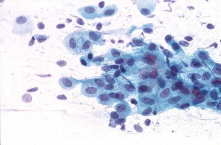
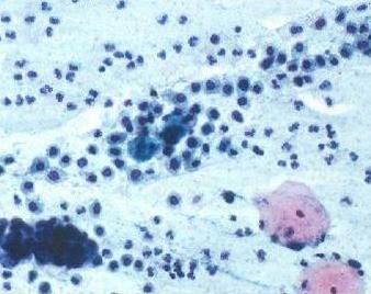
Istiociti, leucociti e globuli rossi
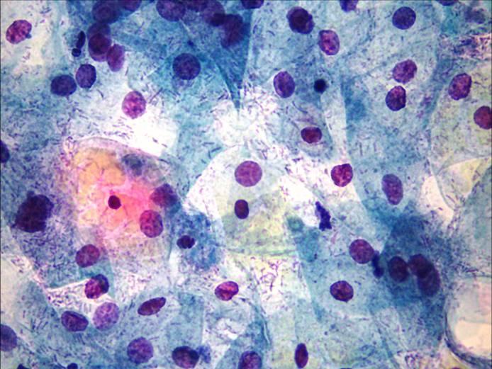
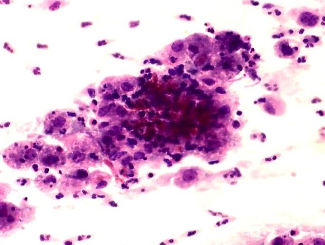
HISTIOCYTES / MACROPHAGES These delicate cells (10-12 u) have foamy cytoplasm with ill defined cellular borders and eccentric bean shaped, oval or rounded nuclei. The cytoplasm sometimes contains ingested intracytoplasmic material. They are always dissociated and when occuring singly may be difficult to distinguish from discrete glandular cells.They are often found in large aggregates at the end of menstruation (the exodus). Multinucleate giant histiocytes may be found in smears from women with chronic cervicitis and in post-menopausal women.Histiocytes: note oval bean shaped or rounded eccentric nuclei and foamy cytoplasm. Note also size of histiocytes relative to the occasional polymorph or lymphocyte in this field.Histiocytes: note oval bean shaped or rounded eccentric nuclei and foamy cytoplasm. in this aggregate of cellsSpray of red blood cells
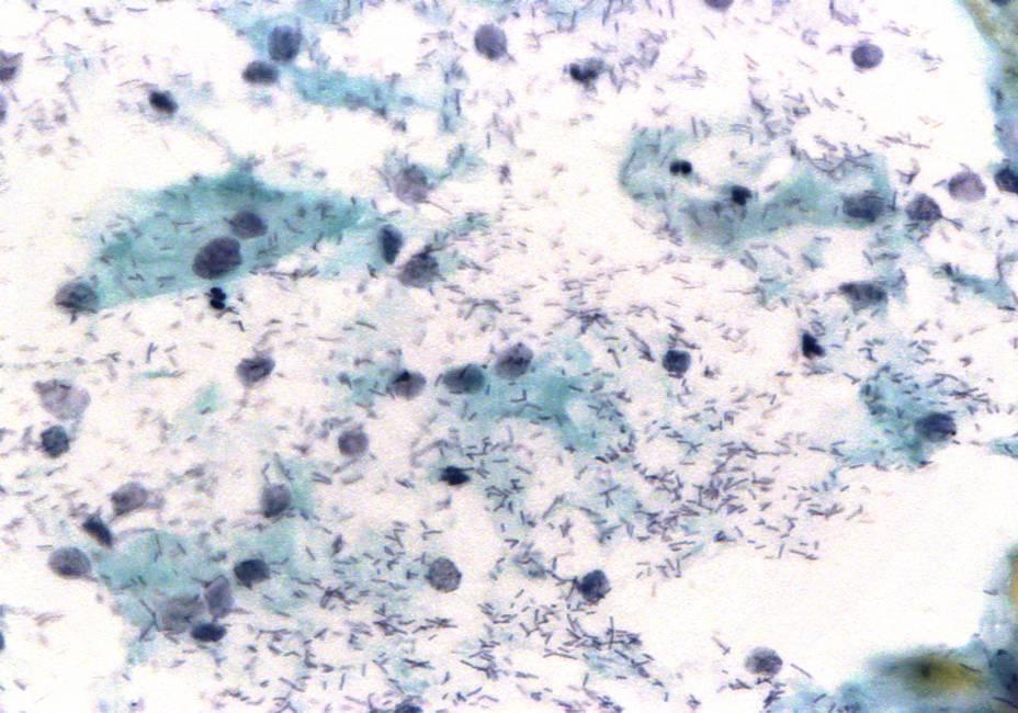
La flora normalmente presente nella vagina (lacto bacilli, gardnerella vaginalis, leptothrix vaginalis)
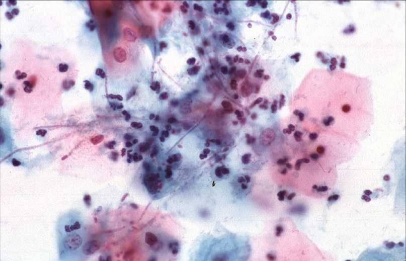
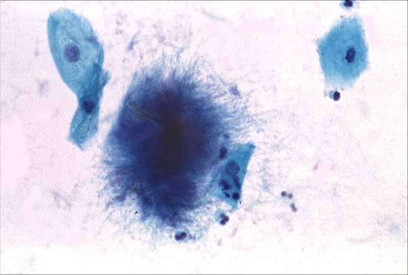
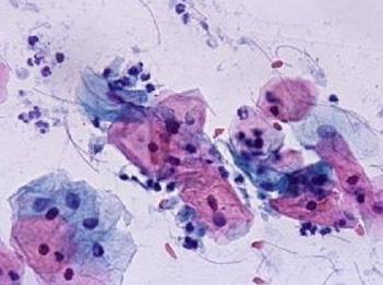
GARDNERELLA VAGINALIS is a small Gram –negative club shaped organisms which stains dark blue with the Papanicolaou stain giving a smear a “dirty” appearance. They accumulate on the surface of large squames to produce the so called “clue cells”. G. vaginalis can be isolated from the genital tract in 20% of asymptomatic women but is also linked to the pathological condition known as bacterial vaginosis. Note absence of inflammatory response in the above smear.GARDNERELLA VAGINALIS is a small Gram –negative club shaped organisms which stains dark blue with the Papanicolaou stain giving a smear a “dirty” appearance. They accumulate on the surface of large squames to produce the so called “clue cells”. G. vaginalis can be isolated from the genital tract in 20% of asymptomatic women but is also linked to the pathological condition known as bacterial vaginosis. Note absence of inflammatory response in the above smear.DODERLEIN BACILLI: These lactobacili appear as blue staining rods 1-2 ?m in length in the background to the smear. They metabolise the glycogen in the intermediate cells and maintain the acid pH of the vagina. They are particularly abundant in smears from pregnant women. This results in cytolysis with destruction of the cytoplasm. The intermediate cells appear ragged and bare intermediate cells nuclei are commonly seen. This pattern is sometimes seen in women on oral contraception and in the late secretory phase of the cycle. Cytolysis can be so marked as to render a smear unsuitable for evaluation.CANDIDA spp: This yeast like fungus is found in the genital tract of 20% of pregnant women. It is also found in diabetics and immunosuppressed patients. The yeast and psueomycelium forms a mesh over the surface of the squamous cells, the pseudohyphe and spores staining faintly with the Pap stain. Most often the woman is symptomless but infection is also associated with an irritating white discharge.ACTINOMYCES; This group of bacteria is frequently found in women fitted with an intrauterine contraceptive device. Morphologically it appears As a fluffy ball of bacteria 20-100Uin diameter with slender branching threads streaming from it. Normally inflammatory reaction is minimal and the organism causes no symptoms although there is a small risk of ascending infection. Non pathogenic amoeba, Entamoeba gingivalis have been found in association with actinomycosis.LEPTOTHRIX VAGINALIS are non pathogenic organisms which are easily recognised in cervical smears. They are apparent as long threads up to 40u In length. They are frequently found in association with Trichomonas vaginalis.
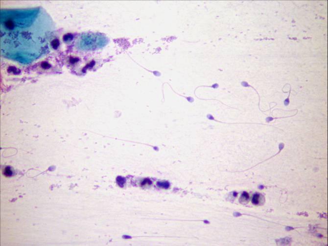
Contaminanti es spermatozoi, granuli di talco
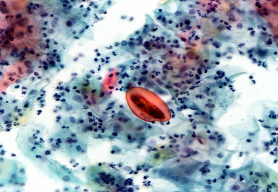
Spermatozoa and spermatocytes – There are numerous spermatozoa in the background of this post coital smear.Spermatozoa and spermatocytes – Note spermatozoa, spermatocytes and amorphous debris in the background of this smear.Talc granules – CONTAMINATING TALC GRANULES: These contaminants are derived from the talc on the gloves used by the smear taker. They stain a variety of colours with the Pap stain but can be recognised by their characteristic shape and appearance under refracted light.Talc granules – CONTAMINATING TALC GRANULES: These contaminants are derived from the talc on the gloves used by the smear taker. They stain a variety of colours with the Pap stain but can be recognised by their characteristic shape and appearance under refracted light.Airdrying artifact – CORNFLAKE ARTIFACT: This common brown artifact is said to be caused by air bubbles formed when xylene dries before the slide is mounted. It can sometimes be so extensive as to render the slide unsuitable for evaluation. Remounting the slide can sometimes improve the appearance of the smear.Haematoidin crystals (cockleburrs) – These reddish brown crystalline arrays are formed from bile like pigment and usually indicate old bleeding.Ovum of enterobius vermicularis in cervical smear – an ovoid shell 20- 60um which is flattened on one side; a faecal contaminant Adult worms are occasionally found.
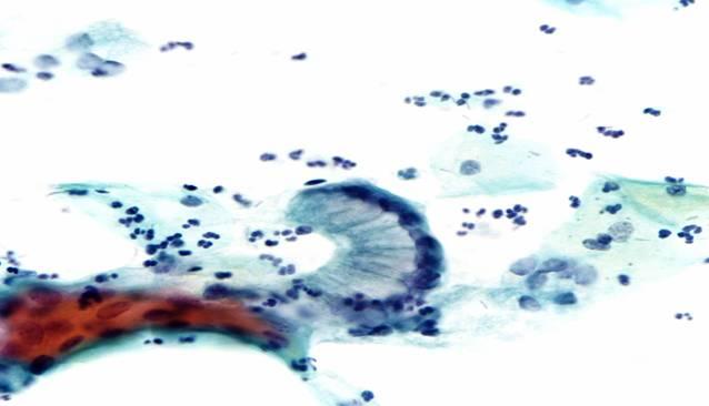
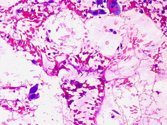
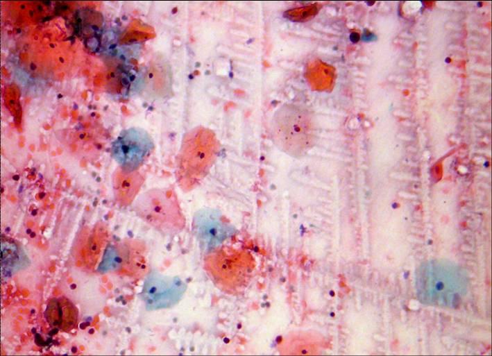
Lembi di muco cervicale
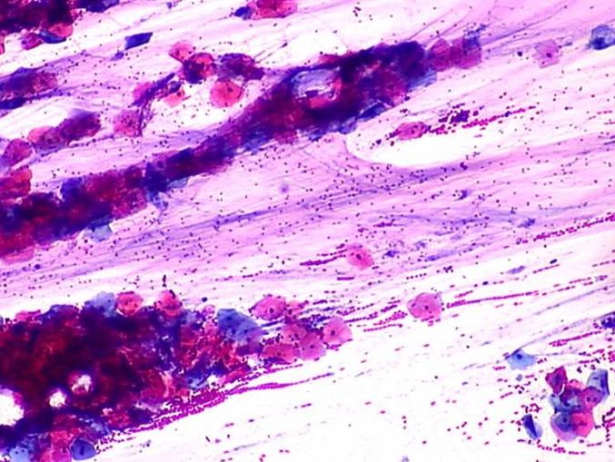
Glandular cells of endocervical origin – Ferning can sometimes be seen mid cycle when the cervical mucous forms a find miscellar network to facilitate the passage of spermatozoa.
Manage Cookie Consent
To provide the best experiences, we use technologies like cookies to store and/or access device information. Consenting to these technologies will allow us to process data such as browsing behavior or unique IDs on this site. Not consenting or withdrawing consent, may adversely affect certain features and functions.
Functional
Sempre attivo
The technical storage or access is strictly necessary for the legitimate purpose of enabling the use of a specific service explicitly requested by the subscriber or user, or for the sole purpose of carrying out the transmission of a communication over an electronic communications network.
Preferences
The technical storage or access is necessary for the legitimate purpose of storing preferences that are not requested by the subscriber or user.
Statistics
The technical storage or access that is used exclusively for statistical purposes.The technical storage or access that is used exclusively for anonymous statistical purposes. Without a subpoena, voluntary compliance on the part of your Internet Service Provider, or additional records from a third party, information stored or retrieved for this purpose alone cannot usually be used to identify you.
Marketing
The technical storage or access is required to create user profiles to send advertising, or to track the user on a website or across several websites for similar marketing purposes.