

Evaluation of the specimen
Several features will be considered in the evaluation of the specimen: the type of cells (such as thyrocytes, macrophages, lymphocytes) Thyrocytes and macrophages. Fluid colloid in the background, bland thyrocytes and pigmented macrophages. Lymphocytes and thyrocytes. A group of bland thyroid cells showing a follicular arrangement, surrounded by smaller lymphoid cells with little amount of identifiable cytoplasm. […]
Multinodular goitre
It is a nodular enlargement of the gland, due to derangements in hormone production, which is often asymmetric and sometimes extreme. Follicular cells undergo hyperplasia, leading to the formation of several nodules. The nodules, which are usually not encapsulated, may considerably vary in their microscopic appearance: some of them are composed of very large macrofollicles […]
Papillary carcinoma
Papillary thyroid carcinoma (PTC) is the most common malignancy of the thyroid (>80% of thyroid cancers). It may occur at any age, but most patients are between 30 and 50 years old; most of them are female (F:M 3:1). It usually presents as a solitary, non-functioning (‘cold’) nodule, sometimes with cervical lymphadenopathy due to metastatization. […]
Terminology for reporting results
Terminology for reporting results It is advisable to use diagnostic categories such as negative (benign), suspicious, positive (malignant) and non-diagnostic (unsatisfactory), followed by a description. Negative (benign) aspirate Positive (malignant) aspirate Suspicious aspirate An increasingly used classification identifies five categories basically splitting the suspicious category into probably reactive (T3 – low risk of malignancy) and […]
Radiation changes
Long-term morphologic changes may occur in the thyroid of patients who undergo external irradiation of the neck (usually as a therapy of malignancies) or administration of radioactive iodine (which is used to treat hyperthyroidism). Cytologic diagnostic features sheets (macrofollicles) enlarged cells normal N/C ratio Hürthle cell change cytoplasmic vacuolization marked nuclear atypia: marked size variation […]
Thyroid
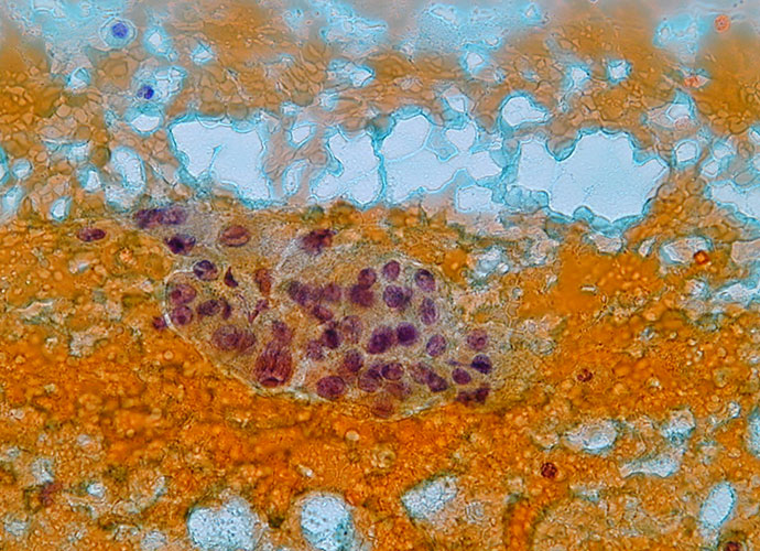
On completion of this section the cytotechnologist should know: the anatomy and histology of the normal thyroid the role of the thyroid hormones and the control of their production the incidence of thyroid nodules and the diseases which can present as thyroid nodules the diagnostic tests in thyroid nodules the indications of thyroid fine needle […]
Anatomy and Physiology
Anatomy The thyroid is an endocrine organ situated in the neck. Its position, close to the thyroid cartilage gave the organ its name as thyroid is derived from the Greek word thyros, meaning shield it was originally considered to protect the larynx. Embryologically the thyroid develops at the base of the tongue from the […]
Thyroid nodules
Thyroid nodules Palpable thyroid nodules affect 4-7% of the adult population with female predominance, although it has been predicted that between 40-70% of adults may have clinically undetected nodules only identifiable by ultrasound or autopsy. The vast majority of these nodules are benign, with only 5-10% proving malignant. Clearly therefore surgical excision of all palpable […]
Classification of thyroid neoplasms
WHO classification of thyroid tumours (1988) I. Epithelial tumours Benign Follicular adenoma and variants Others including hyalinizing trabecular adenoma Malignant Follicular carcinoma including minimally invasive, widely invasive and poorly differentiated subtypes Papillary carcinoma and variants including papillary microcarcinoma, follicular variant and diffuse sclerosing variant Medullary carcinoma and variants including mixed medullary-follicular carcinoma Undifferentiated (anaplastic) carcinoma […]
Fine needle aspiration (FNA)
Diagnostic tests in thyroid nodules Clinical history and examination are the first step to approach the investigation of the thyroid nodule; however they are not sufficiently accurate by themselves. Ultrasound examination, which can discriminate between a solid and a cystic nodule, does not reliably predict malignancy. Radionuclide imaging is not useful to identify malignant lesions: […]