Le modificazioni infiammatorie nella cervice descritte sopra sono comuni in tutti I casi di cerviciti acute. In alcuni casi l’effettivo organismo microbiologico causa della cervicite può essere riconosciuto nello striscio o può causare alterazioni citopatiche caratteristiche che permettono la diagnosi di una specifica infiammazione.
Una lista delle specifiche infiammazioni identificate dalla citologia :
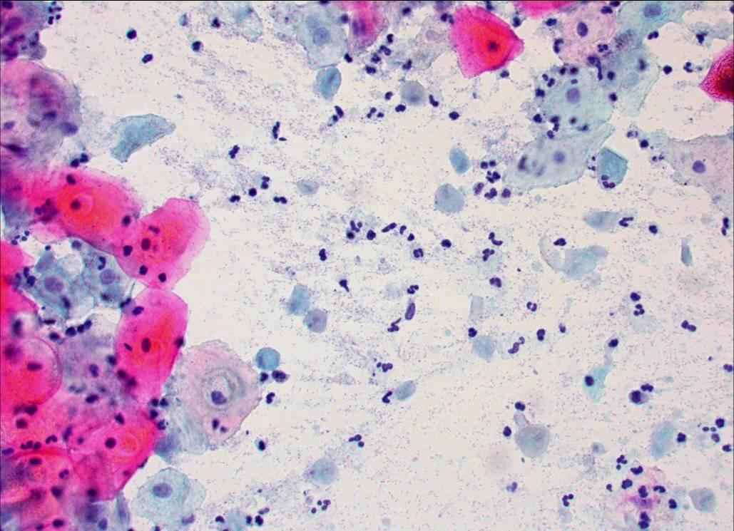
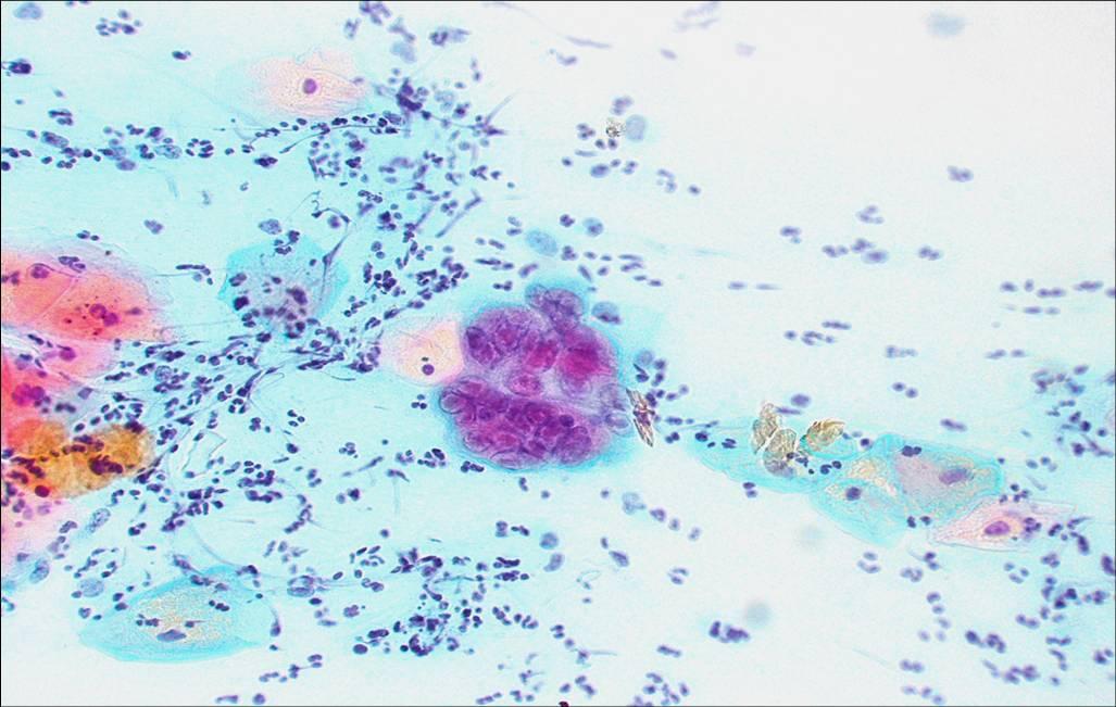
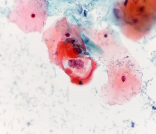
Trichomonas vaginalis
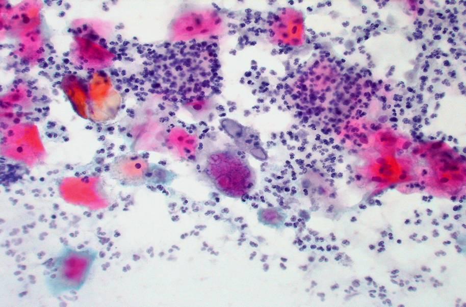
Candida sp
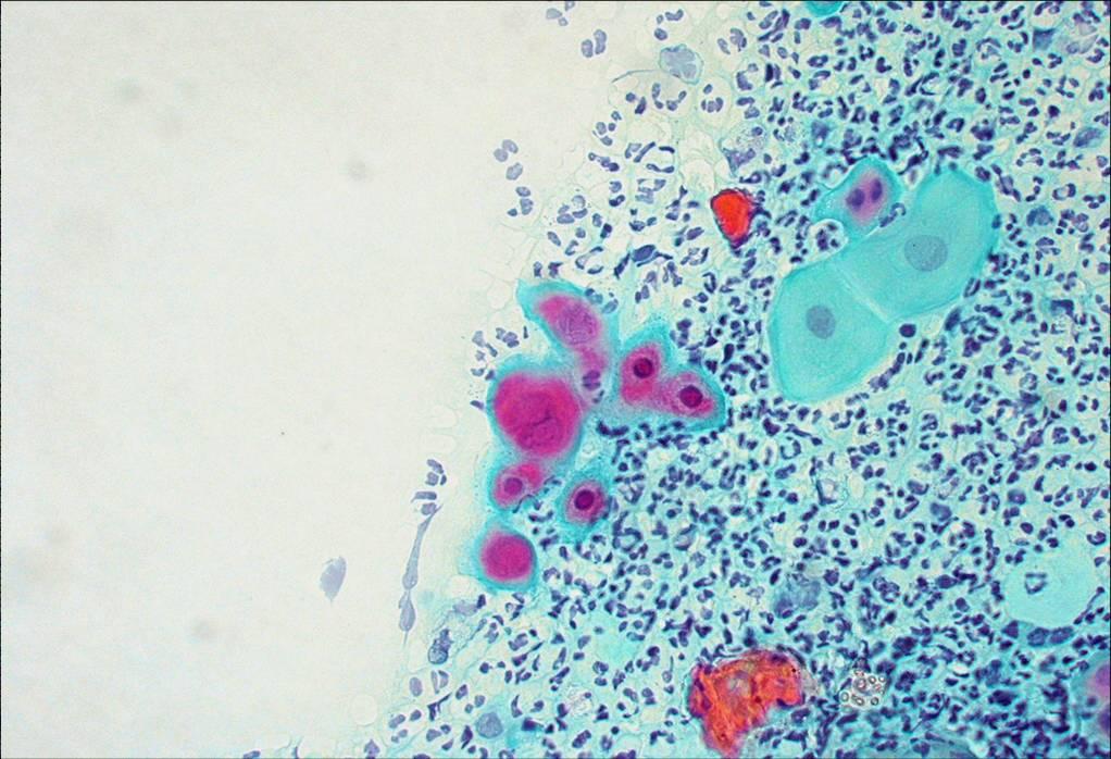
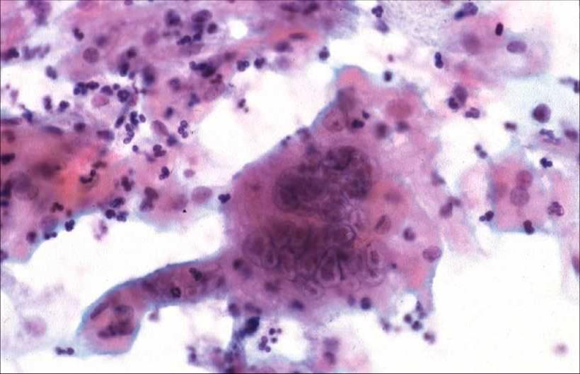
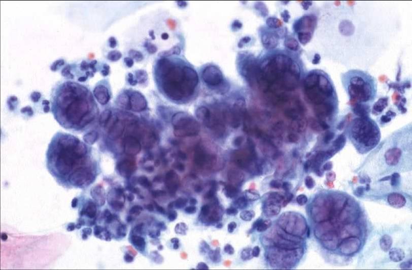
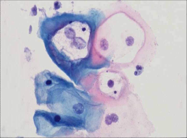
Herpes genitalis
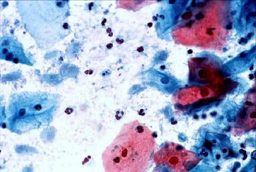
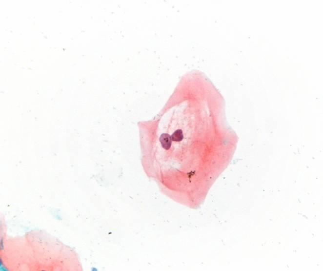
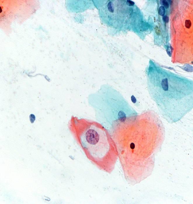
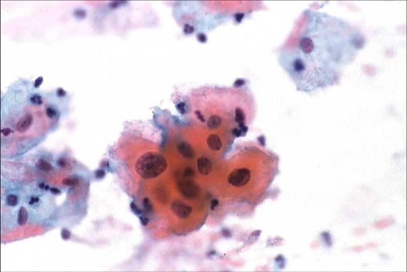
Human Papillomavirus
Actinomyces sp
Chlamydia trachomatis
Infezioni da parassiti come with Schistosoma haematobium , S. mansoni and Fliaria bancrofti possono essere riconosciuti nello striscio cervicale.Possono poi essere identificate lesioni granulomatose suggestive di tubercolosi o inclusioni citomegaliche.
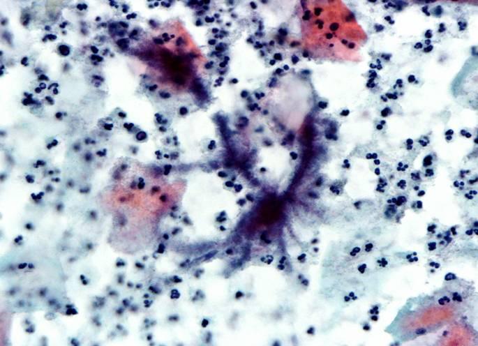
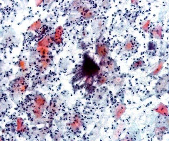
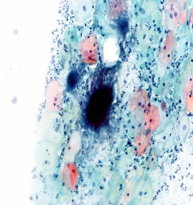
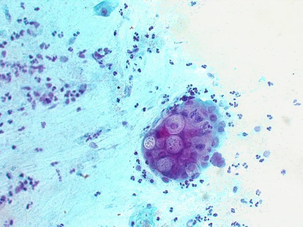
Trichomonas vaginalis: Trichomonads are cyanophilic. flagellate protozoa, shape like a pear, varying in diameter from 15 to 30 microns. The nucleus is vesicular, pale, eccentrically located, and the cytoplasm. Sometimes contains eosinophilic granules. Leptothrix may be seen in association with T. Vaginalis; this finding alone is not diagnostic, but suggests the presence of trichomonads. They are seen here in a coccal background. Smears containing trichomonads often contain highly eosinphilic mature squamous cells and the nuclei of the squames are sometimes surrounded by a small perinuclear halo.Trichomonas vaginalis: Trichomonads are cyanophilic flagellate protozoa, shape like a pear, varying in diameter from 15 to 30 microns. The nucleus is vesicular, pale, eccentrically located, and the cytoplasm. Sometimes contains eosinophilic granules. They are seen here in a coccal background. Note inflammatory backgroundof debris and polymorphs. Clinically infection is characterised by offensive copious yellow /green vaginal discharge.Candida sp. These yeasts are normally commensal in the vagina but may become pathogenic if the pH of the vagina changes as in pregnancy. Infection is common in immunocompromised patients. The yeasts are 3-7 microns in length. They appear gray-brown or eosinophilic with the Papanicolaou stain. Pseudohyphae are formed by elongated budding of the yeast cells and show constrictions. Culture is necessary to distinguish species. Infection is characterised clinically by white itchy discharge and white patches on vagina and cervical mucosa.Histological section of cervix showing acute herpes cervicitis. Note multi nucleate giant cell in the epithelial layer, dissociation of the epithelial layers and coagulative necrosis of the surface epithelial cells. There is infiltration of the epithelium with polymorphs and hyperaemia of the blood vessels.Herpes Simplex Virus. The cytopathic changes of herpes infection can be recognised in this smear. There are two multi nucleate giant cells with the individual nuclei showing a “ground-glass” appearance with a well defined nuclear membrane caused by peripheral margination of chromatin. Other epithelial cell nuclei are showing coagulative necrosis. Note the heavy inflammatory infiltrate.Herpes Simplex Virus. The cytopathic changes of herpes infection can be recognised in epithelial cell nuclei which assume a “ground-glass” appearance with a well defined nuclear membrane caused by peripheral margination of chromatin. The nucleus occasionally contains an eosinophilic intranuclear inclusion. A large large multinucleate giant cell can be seen in the center of the field. Note degenerative changes of the epithelial cells and inflammatory cells in the background.Herpes Simplex Virus. The cytopathic changes of herpes infection can be recognised in the epithelial cell nuclei which assume a “ground-glass” appearance with a well defined nuclear membrane caused by peripheral margination of chromatin. A large large multinucleate giant cell can be seen in the center of the field. The nuclei contain large type A intranuclear inclusions.Herpes Simplex Virus. A high power view of multinucleate giant cell showing the large type A nuclear inclusions. It is said that inclusions are found in the late stages of the infection.Herpes Simplex Virus. A high power view of multinucleate giant cells showing ground glass appearance of the nuclei and margination of the chromatin. Moulding of the nuclei is a characteristic feature of herpes infected multinucleate cells.Human papillomavirus infection is characterised in cervical smears by the presence of koilocytes which are pathognominic for this infection. Koilocytes are mature squamous cells with a characterisitic clear halo surrounding the nucleus. The halo is well defined and surrounded by a sharp edge of eosinophilic cytoplasm. The nuclei may be multiple and are slightly enlarged reflecting viral replication. According to the Bethesda System smears containing koilocytes are classified as ASC-US or LSIL.Human papillomavirus infection is characterised in cervical smears by the presence of koilocytes which are pathognominic for this infection. Koilocytes are mature squamous cells with a characterisitic clear halo surrounding the nucleus. The halo is well defined and surrounded by a sharp edge of eosinophilic cytoplasm. The nuclei may be multiple and are slightly enlarged reflecting viral replication. According to the Bethesda System smears containing koilocytes are classified as ASC-US or LSIL.Human papillomavirus infection is characterised in cervical smears by the presence of koilocytes which are pathognominic for this infection. Koilocytes are mature squamous cells with a characterisitic clear halo surrounding the nucleus. The halo is well defined and surrounded by a sharp edge of eosinophilic cytoplasm. The nuclei may be multiple and are slightly enlarged reflecting viral replication. According to the Bethesda System smears containing koilocytes are classified as ASC-US or LSIL.Human papillomavirus infection is characterised in cervical smears by the presence of koilocytes which are pathognominic for this infection. Koilocytes are mature squamous cells with a characterisitic clear halo surrounding the nucleus. The halo is well defined and surrounded by a sharp edge of eosinophilic cytoplasm. The nuclei may be multiple and are slightly enlarged reflecting viral replication. According to the Bethesda System smears containing koilocytes are classified as ASC-US or LSIL.Individual small keratinised squamous cells with slightly atypical nuclei (dyskeratocytes) are also found in smear in the presence of HPV infection. They reflect the abnormal keratinisation associated with this infection. They are, by convention, classified as ASC-US or LSIL. ACTINOMYCES; This group of anaerobic gram positive bacteria is frequently found in women fitted with an intrauterine contraceptive device. Morphologically it appears as a fluffy ball of bacteria 20-100Uin diameter with slender branching threads streaming from it. Normally inflammatory reaction is minimal and the organism causes no symptoms. It can rarely cause acute cervicitis,ascending infection and infertility. Note the inflammatory response in this smear. It is usual to remove the IUD if the patient complains of dysmenorrhoea or discharge.ACTINOMYCES; This group of bacteria is frequently found in women fitted with an intrauterine contraceptive device . Morphologically it appears as a fluffy ball of bacteria 20-100Uin diameter with slender branching threads streaming from it. Normally inflammatory reaction is minimal and the organism causes no symptoms. It can rarely cause acute cervicitis, ascending infection and infertility. Note the inflammatory response in this smear. It is usual to remove the IUD if the patient complains of dysmenorrhoea or discharge. ACTINOMYCES; This group of bacteria is frequently found in women fitted with an intrauterine contraceptive device . Morphologically it appears as a fluffy ball of bacteria 20-100Uin diameter with slender branching threads streaming from it. Normally inflammatory reaction is minimal and the organism causes no symptoms. It can rarely cause acute cervicitis, ascending infection and infertility. Note the inflammatory response in this smear. It is usual to remove the IUD if the patient complains of dysmenorrhoea or discharge.Chlamydia trachomatis is an obligate intracellular parasite which is associated with the formation of granular intracytoplasmic inclusions. It is a common cause of non specific urethritis and cervicitis and accounts for 20% of all cases of pelvic inflammatory disease and infertility. The cytoplasmic inclusions can be seen occasionally in immature metaplastic cells but cytological diagnosis is unreliable and confirmation by immunofluorescence is essential. Follicular cervicitis may be present.Chlamydia trachomatis is a small intracellular obligate bacterium which is associated with the formation of granular intracytoplasmic inclusions. It is a common cause of non specific urethritis and cervicitis and accounts for 20% of all cases of pelvic inflammatory disease and infertility. The cytoplasmic inclusions can be seen occasionally in immature metaplastic cells but cytological diagnosis is unreliable and confirmation by immunofluorescence is essential. Follicular cervicitis may be present.
Manage Cookie Consent
To provide the best experiences, we use technologies like cookies to store and/or access device information. Consenting to these technologies will allow us to process data such as browsing behavior or unique IDs on this site. Not consenting or withdrawing consent, may adversely affect certain features and functions.
Functional
Sempre attivo
The technical storage or access is strictly necessary for the legitimate purpose of enabling the use of a specific service explicitly requested by the subscriber or user, or for the sole purpose of carrying out the transmission of a communication over an electronic communications network.
Preferences
The technical storage or access is necessary for the legitimate purpose of storing preferences that are not requested by the subscriber or user.
Statistics
The technical storage or access that is used exclusively for statistical purposes.The technical storage or access that is used exclusively for anonymous statistical purposes. Without a subpoena, voluntary compliance on the part of your Internet Service Provider, or additional records from a third party, information stored or retrieved for this purpose alone cannot usually be used to identify you.
Marketing
The technical storage or access is required to create user profiles to send advertising, or to track the user on a website or across several websites for similar marketing purposes.