This content is also available in:
![]() English
English ![]() Español
Español
Caratteristiche citologiche e morfologiche della CIN
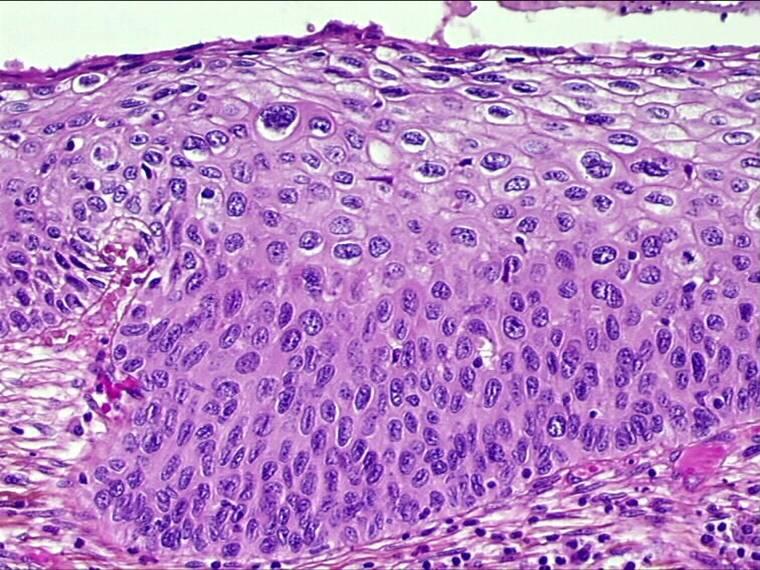
- In tutti i casi di CIN, il normale epitelio della cervice è sostituito da cellule epiteliali atipiche che mostrano vari gradi di differenziazione.
- Cellule neoplastiche o paraneoplastiche indifferenziate di aspetto basaloide o parabasaloide possono occupare l’intero spessore dell’ epitelio (CIN3) o possono differenziarsi man mano procedono verso la superficie (CIN1,CIN2).
- I nuclei delle cellule indifferenziate sono grandi, irregolari, con variazione di forma e volume. Il contenuto in cromatina è aumentato e irregolare nella sua struttura e il rapporto nucleo/citoplasma è aumentato.
- La quantità di citoplasma aumenta quando la cellule neoplastica si differenzia. Irregolarità nucleare, pleomorfismo ed anisonucleosi persistono però anche quando diminuisce la taglia nucleare.
- L’attività mitotica non è più confinata allo strato basale dell’epitelio e mitosi atipiche possono essere viste ad ogni livello.
- E’ poi frequente ritrovare foci di CIN1,CIN2 e CIN3 anche nella stessa cervice.


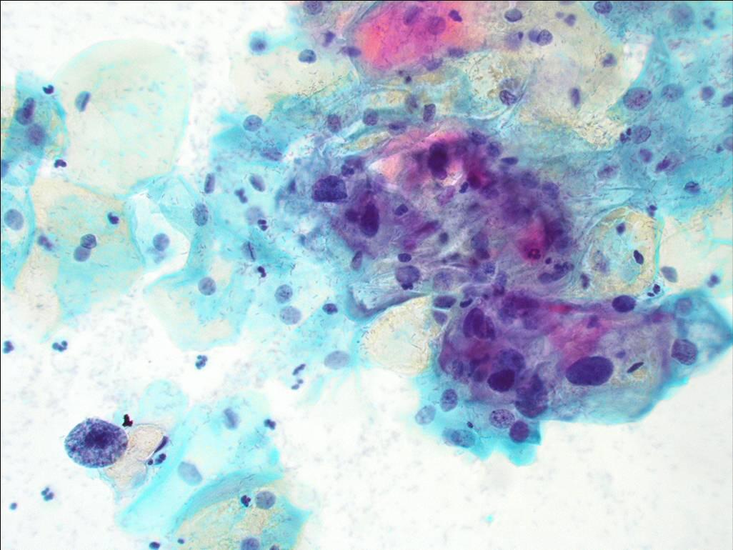
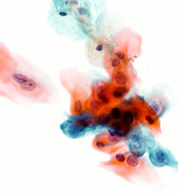
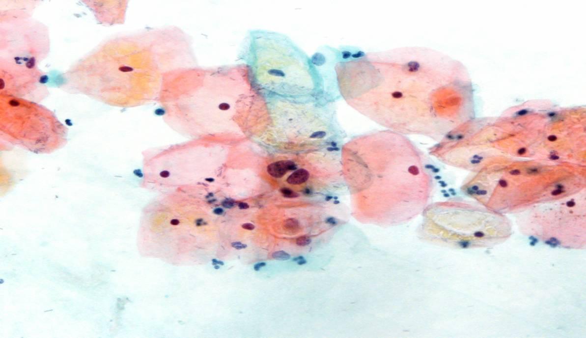
Le cellule atipiche ritrovate nello striscio derivano dalla superficie dei focolai di CIN. Le cellule atipiche possono essere singole in lembi o in foglietti. Possono essere riconosciute rispetto alle cellule normali per le seguenti caratteristiche:
Le caratteristiche morfologiche delle cellule presenti nei foci di neoplasia intraepiteliale cervicale presenti in uno striscio cervicale sono:
- Irregolare ingrandimento nucleare che porta ad un aumento del rapporto nucleo/citoplasma.
- Variazione nella forma e nel volume nucleare.
- Irregolartià nel profilo della membrana nucleare.
- Ipercromasia del nucleo e pattern atipico della cromatina
- Nuclei grandi, irregolari ed a volte multipli (non presenti in CIN1)
- Il grado della CIN (CIN 1, 2 o 3) può essere dedotto dal grado di ingrandimento nucleare e dall’alterazione del rapporto nucleo/citoplasma.
Le tre Tavole ingrandibili sottostanti descrivono e illustrano con serie di diapositive le caratteristiche citologiche e i differenti gradi dellaCIN
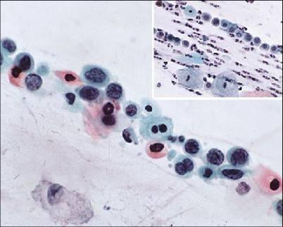
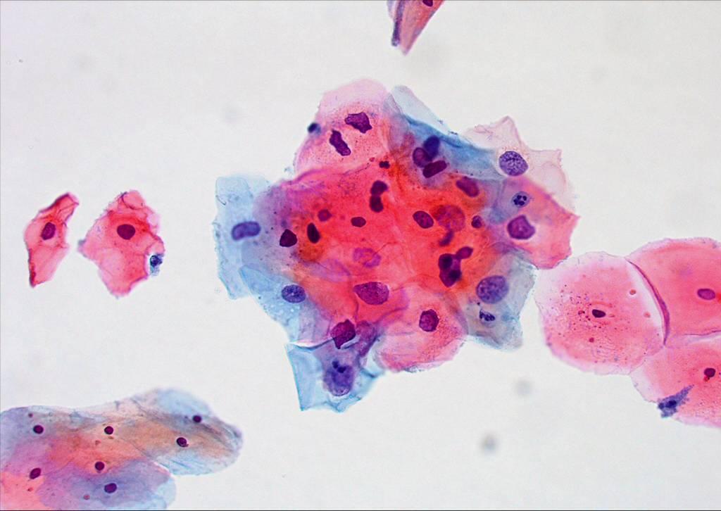
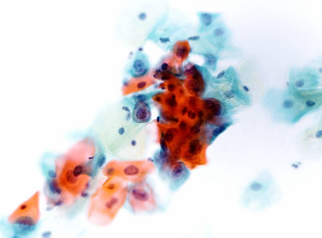
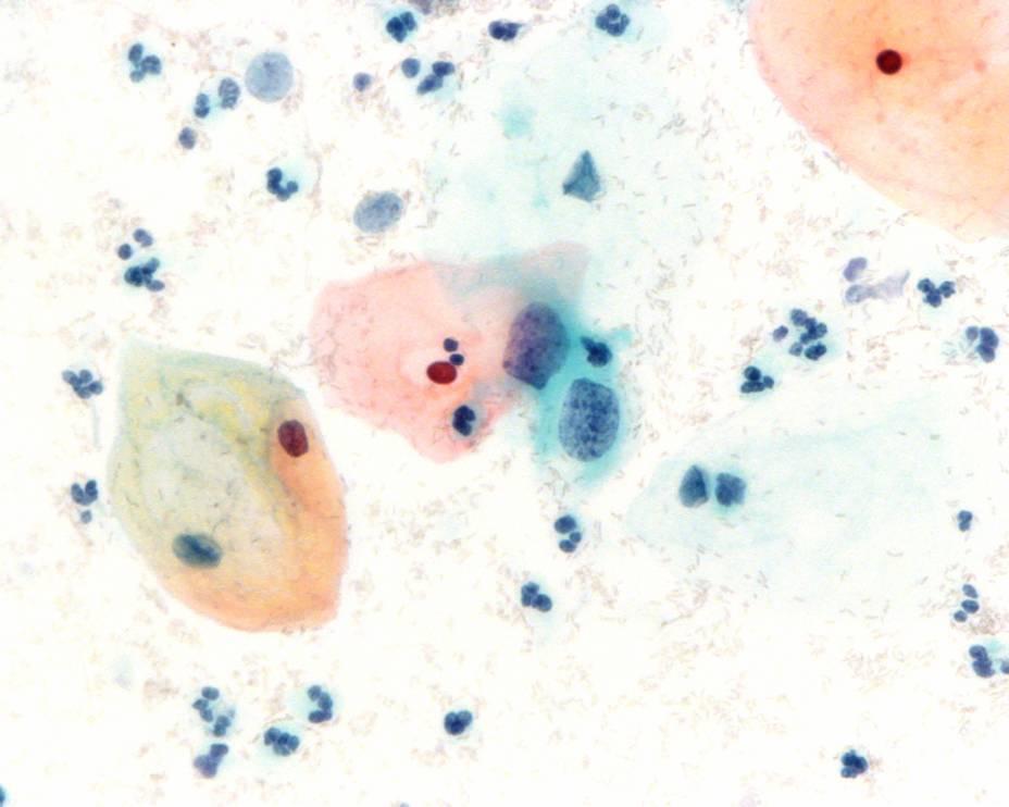
Cytological diagnosis of CIN1 (encompassing LSIL and mild dysplasia)
The Bethesda System definition of CIN 1 (LSIL, mild dysplasia)
- Nuclear enlargement is three or more times that of a normal intermediate cell nucleus*
- Nuclear/cytoplasmic ratio may or may not be altered as cytoplasm is usually abundant and may be keratinised. Cell borders are usually distinct
- Moderate variation in nuclear size and shape
- Binucleation or multinucleation are not uncommon
- Hyperchromasia with uniformly distributed chromatin. The chromatin may appear degenerated or smudged if associated with HPV changes
- Nucleoli are rare or inconspicuous
- Nuclear membrane may or may not be distinct
- Cells with a well-defined, optically empty perinuclear halo and peripheral dense cytoplasm (the so called koilocytotic atypia) must also show the above described nuclear abnormalities to be classified as CIN 1 (LSIL).
*The WHO cytological definition of CIN 1 requires that the nucleus occupies less than one half the area of the cell.

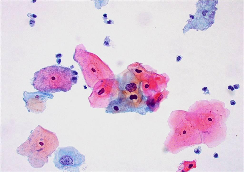
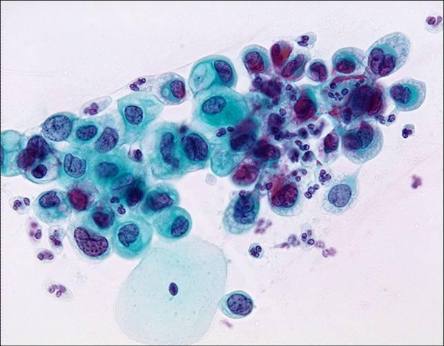
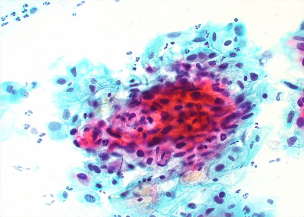
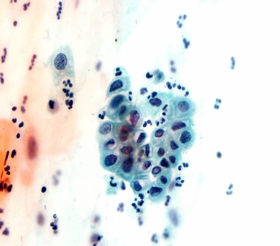
The Bethesda System definition of CIN 2 (HSIL)
The Bethesda System definition of CIN 2 (HSIL)
- Cells occur singly or grouped in sheets or in syncytial-like clusters with crowded nuclei
- Cells generally have delicate or dense cytoplasm; occasionally the cytoplasm is heavily keratinised
- Nuclear enlargement is three or more times greater than that of normal intermediate cell nuclei*
- The cytoplasmic area may be decreased. For this reason, in a cell with very high nuclear/cytoplasmic ratio, the dimension of the nucleus can be smaller than in LSIL
- Nuclear borders are irregular
- Anisonucleosis is prominent
- Hyperchromasia of nuclei is present and chromatin granular & evenly distributed (fine or coarse)
- Overall cell size may be smaller than in LSIL
*The WHO cytological definition of CIN 1 requires that the nucleus occupies between one half and two thirds the area of the cell.
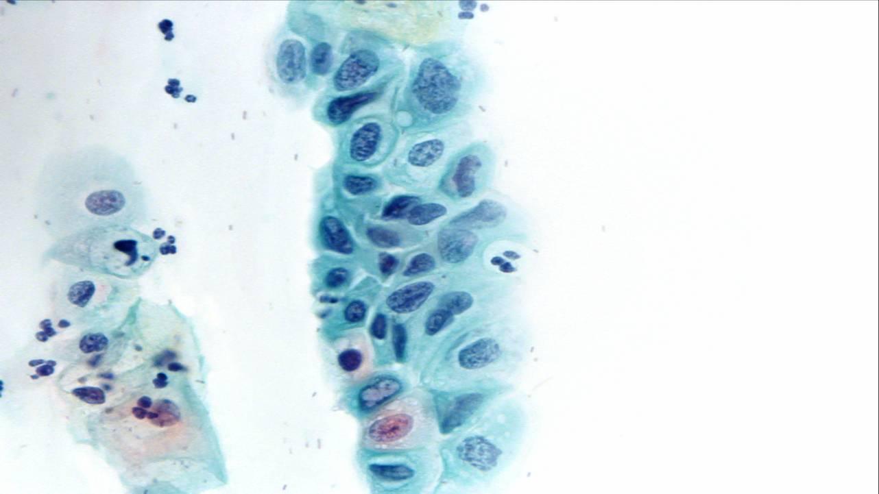
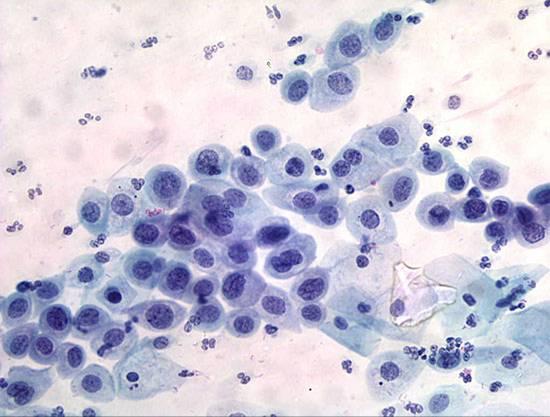
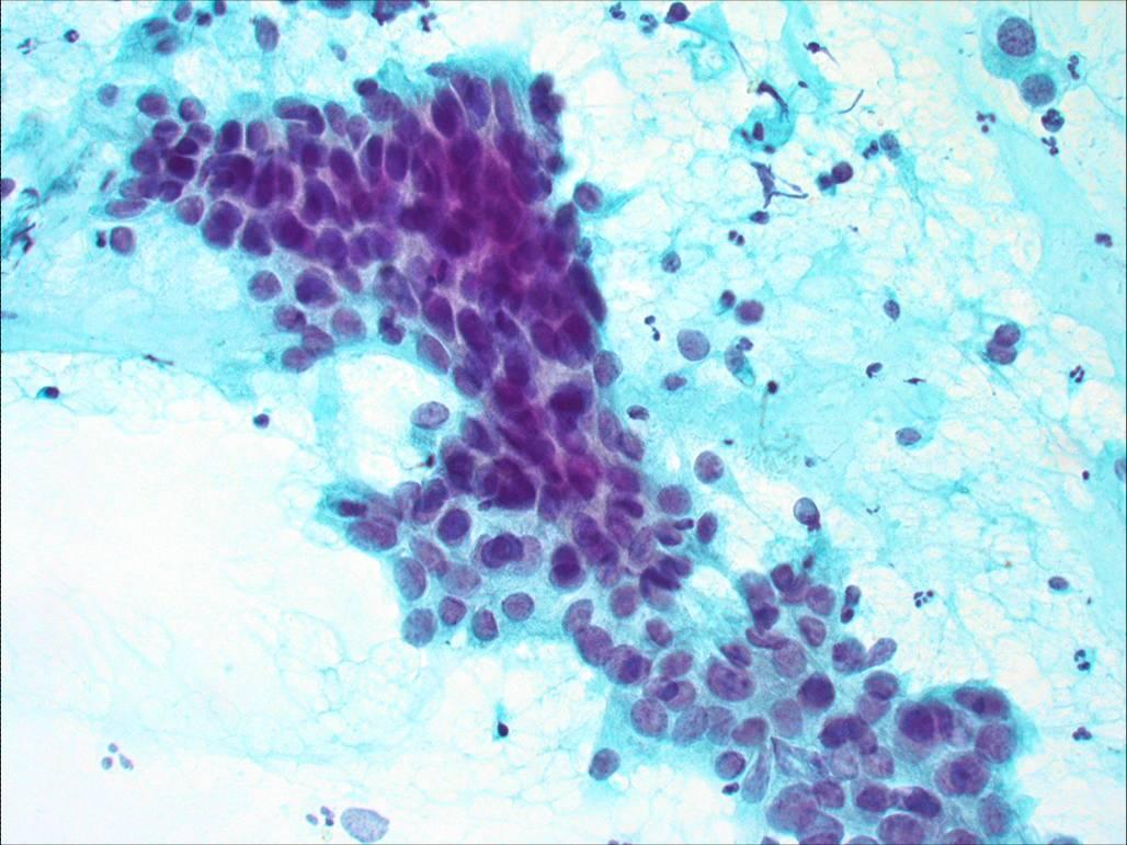
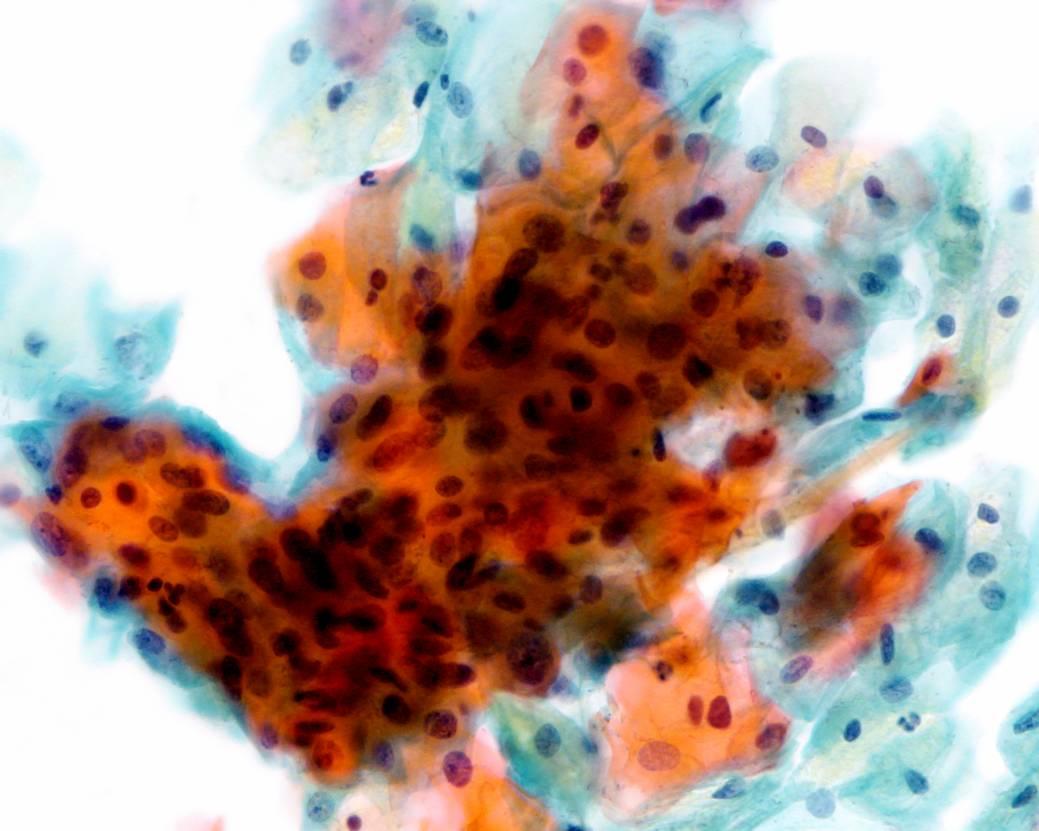
Cytological diagnosis of CIN3 (encompassing HSIL, severe dysplasia and carcinoma in situ)
The Bethesda System definition CIN 3 (HSIL)
- Cells occur singly or grouped in sheets or in syncytial-like clusters
- Abnormal nuclei generally found in cells with “immature”, delicate or dense cytoplasm; occasionally the cytoplasm is heavily keratinised
- Nuclear enlargement is three or more times the area of normal intermediate nuclei*
- The cytoplasmic area is decreased. For this reason, in a cell with very high nuclear/cytoplasmic ratio, the dimension of the nucleus can be smaller than in LSIL
- Overall cell size is often ksmaller than in LSIL
- Nuclei are hyperchromatic usually with granular coarse chromatin
- Nuclear borders are irregular and anisonucleosis is a prominent feature
- When the abnormal cells occur in sheets ,the nuclei are crowded and overlapping and mitotic figures may be seen
*The WHO cytological definition of CIN 3 requires that the nucleus occupies more than two thirds of the area of the cell.