This content is also available in:
![]() Italiano
Italiano ![]() Español
Español ![]() Čeština
Čeština ![]() Magyar
Magyar ![]() Polski
Polski
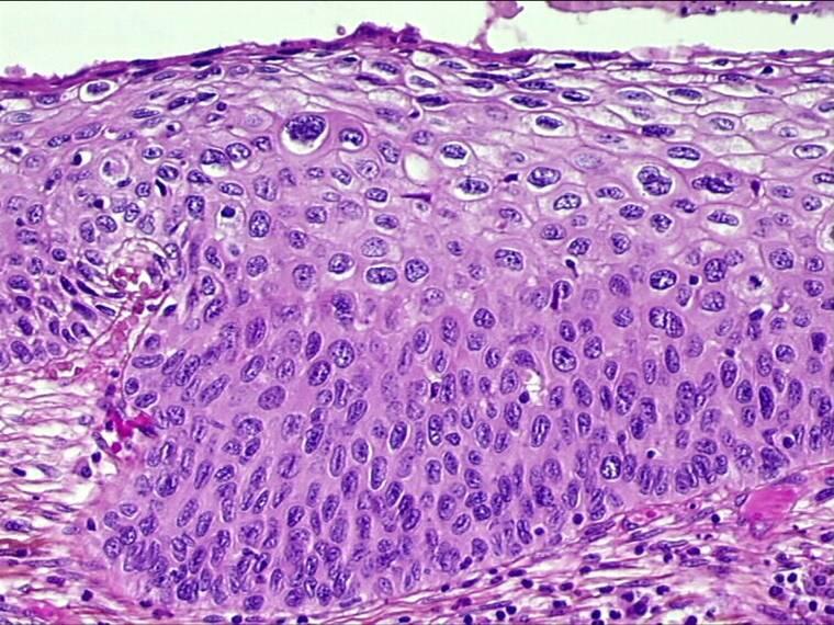
Morphological & biological characteristics of CIN
- In all cases of CIN, the normal epithelium of the cervix is replaced by abnormal epithelial cells showing varying degrees of differentiation.
- Undifferentiated neoplastic cells of basaloid or parabasaloid appearance may occupy the whole thickness of the epithelium (CIN3) or they may differentiate as they approach they approach the surface (CIN1, CIN2)
- The nuclei of the undifferentiated cells are large, irregular and vary in size and shape. The chromatin content is increased and abnormal in structure and the nuclear/cytoplasmic ratio is high.
- As neoplastic cells differentiate the amount of cytoplasm increases. Nuclear irregularity, pleomorphism and anisonucleosis persist although there is usually a decrease in nuclear size.
- Mitotic activity is no longer confined to the basal layers of the epithelium and atypical mitoses may be seen at any level
- It is not uncommon for foci of CIN1, CIN2 and CIN3 to be present in the same cervix


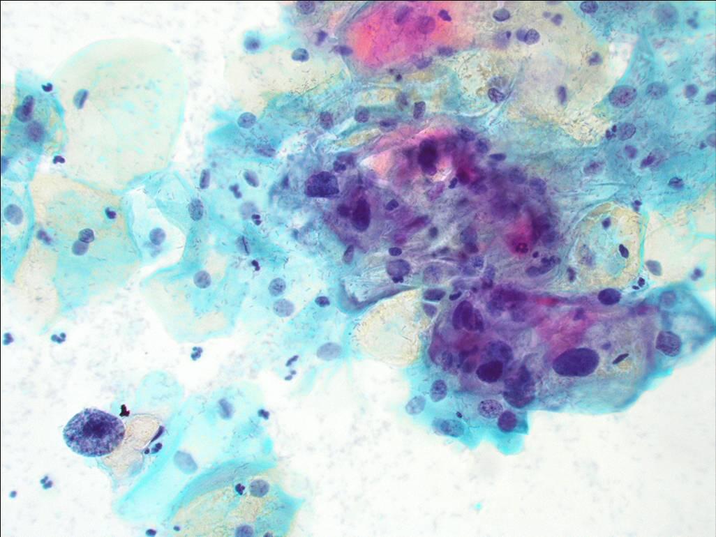
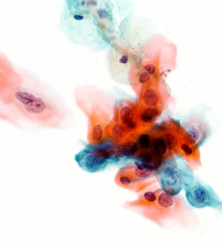
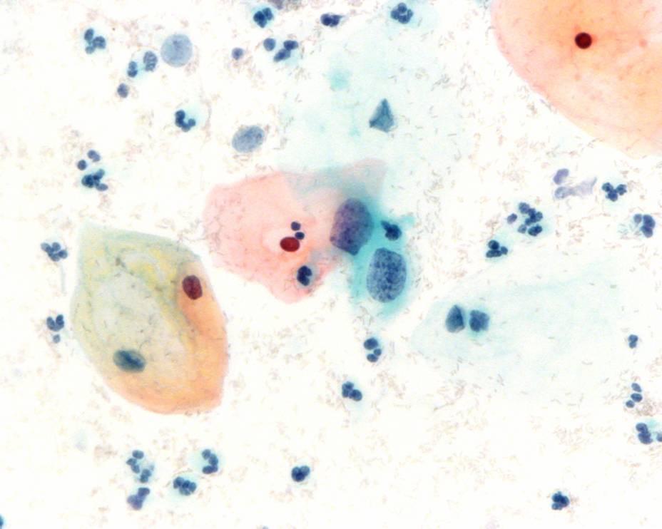
The abnormal cells found in the smear are derived from the surface of the CIN lesion. The abnormal cells appear singly or in streaks or in sheets. They can be distinguished from normal cells by the following features:
The morphology of cells from a focus of cervical intraepithelial neoplasia as they appear in cervical smears
- Disproportionate nuclear enlargement resulting in alteration of normal nuclear/cytoplasmic ratio
- Variation in nuclear size and shape
- Irregularity of nuclear outline
- Hyperchromasia of nucleus and abnormal chromatin pattern
- Large irregular and sometimes multiple nuclei (not in CIN1)
- The grade of CIN (CIN 1, 2 or 3) can be deduced from the degree of nuclear enlargement and alteration of n/c ratio
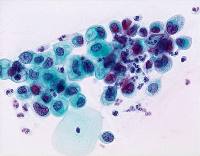
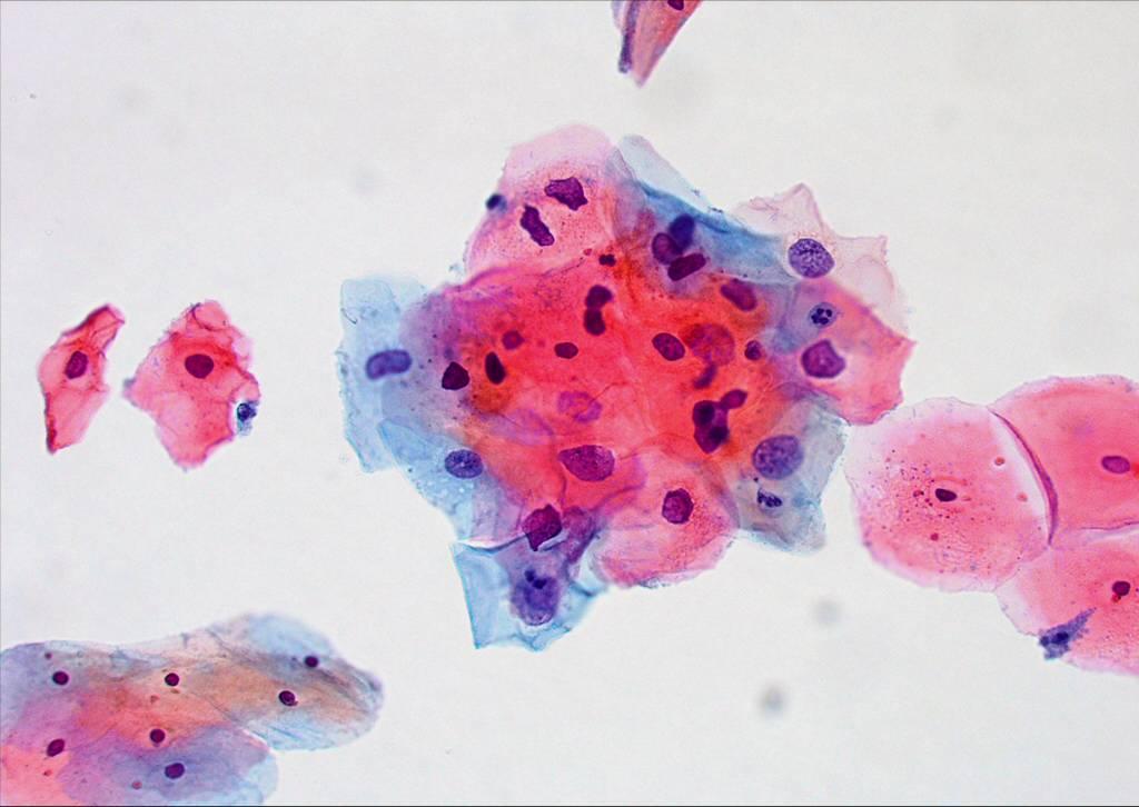
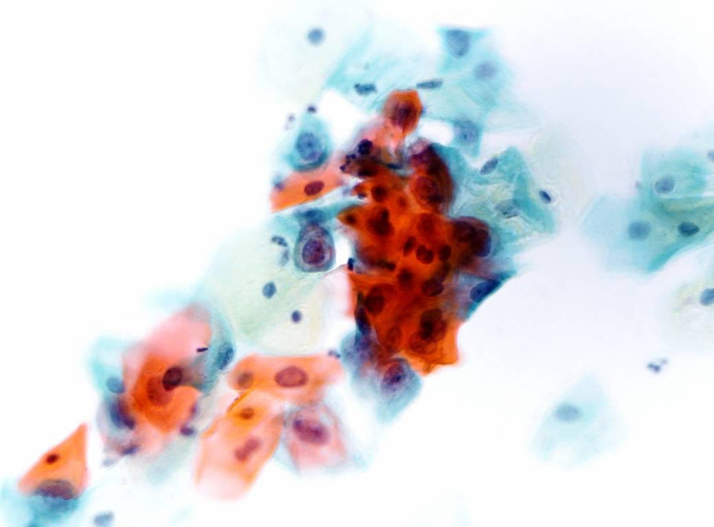
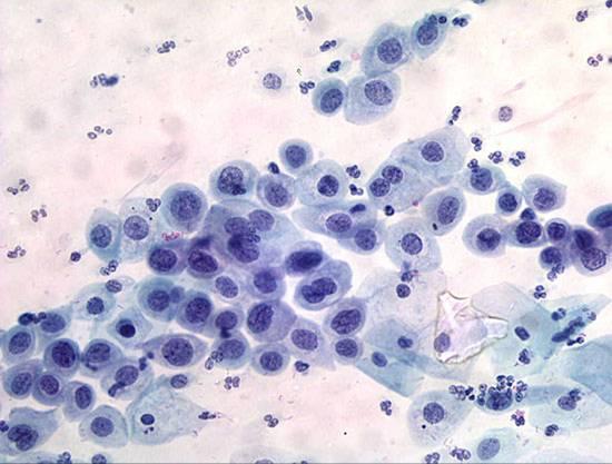
Cytological diagnosis of CIN1 (encompassing LSIL and mild dysplasia)
The Bethesda System definition of CIN 1 (LSIL, mild dysplasia)
- Nuclear enlargement is three or more times that of a normal intermediate cell nucleus*
- Nuclear/cytoplasmic ratio may or may not be altered as cytoplasm is usually abundant and may be keratinised. Cell borders are usually distinct
- Moderate variation in nuclear size and shape
- Binucleation or multinucleation are not uncommon
- Hyperchromasia with uniformly distributed chromatin. The chromatin may appear degenerated or smudged if associated with HPV changes
- Nucleoli are rare or inconspicuous
- Nuclear membrane may or may not be distinct
- Cells with a well-defined, optically empty perinuclear halo and peripheral dense cytoplasm (the so called koilocytotic atypia) must also show the above described nuclear abnormalities to be classified as CIN 1 (LSIL).
*The WHO cytological definition of CIN 1 requires that the nucleus occupies less than one half the area of the cell.

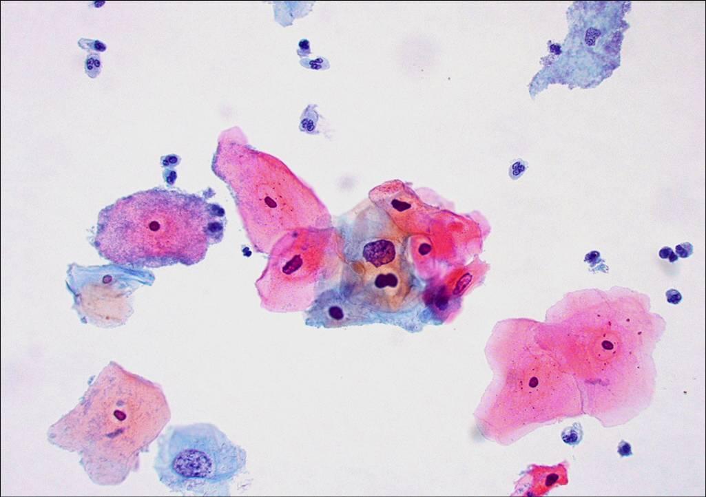
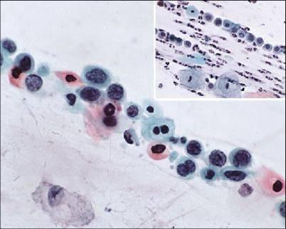
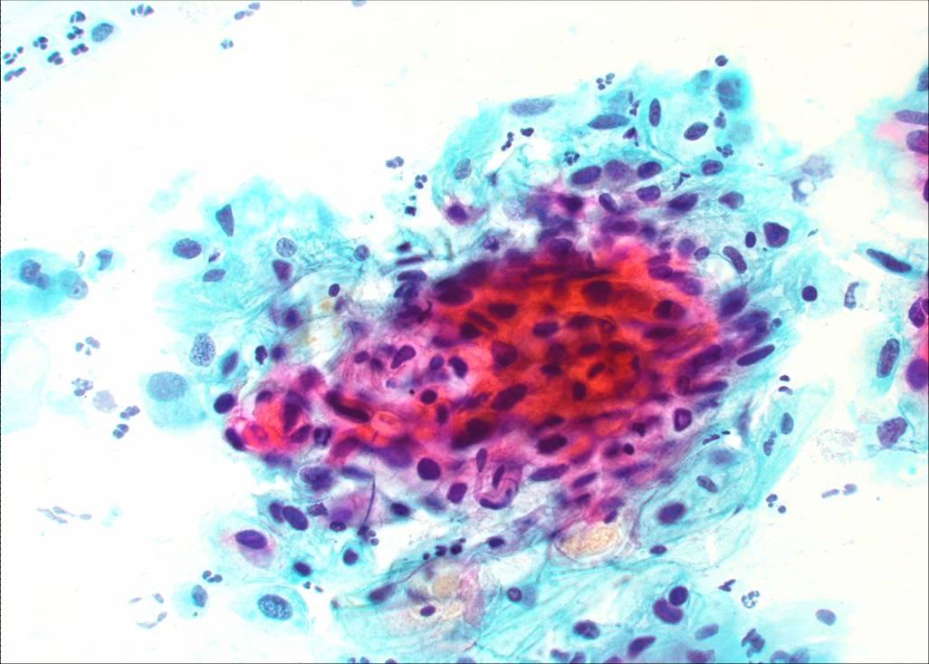
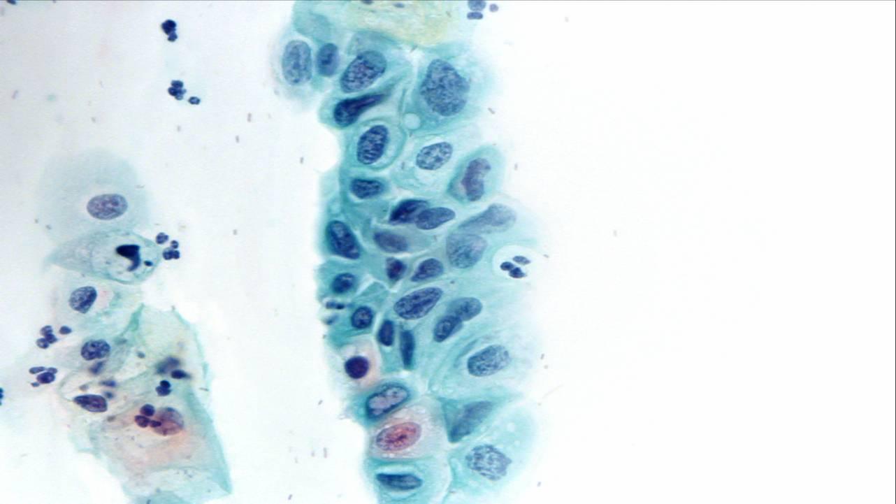
The Bethesda System definition of CIN 2 (HSIL)
The Bethesda System definition of CIN 2 (HSIL)
- Cells occur singly or grouped in sheets or in syncytial-like clusters with crowded nuclei
- Cells generally have delicate or dense cytoplasm; occasionally the cytoplasm is heavily keratinised
- Nuclear enlargement is three or more times greater than that of normal intermediate cell nuclei*
- The cytoplasmic area may be decreased. For this reason, in a cell with very high nuclear/cytoplasmic ratio, the dimension of the nucleus can be smaller than in LSIL
- Nuclear borders are irregular
- Anisonucleosis is prominent
- Hyperchromasia of nuclei is present and chromatin granular & evenly distributed (fine or coarse)
- Overall cell size may be smaller than in LSIL
*The WHO cytological definition of CIN 1 requires that the nucleus occupies between one half and two thirds the area of the cell.
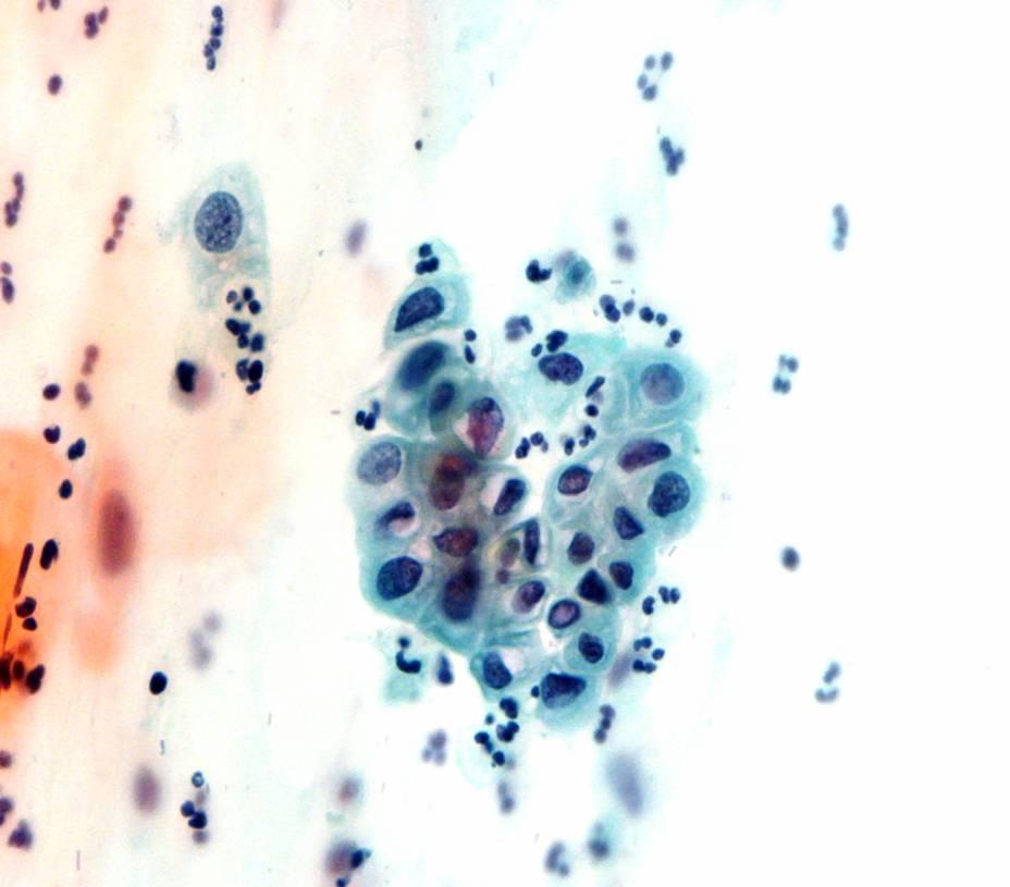
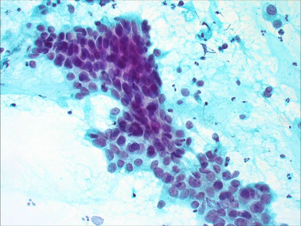
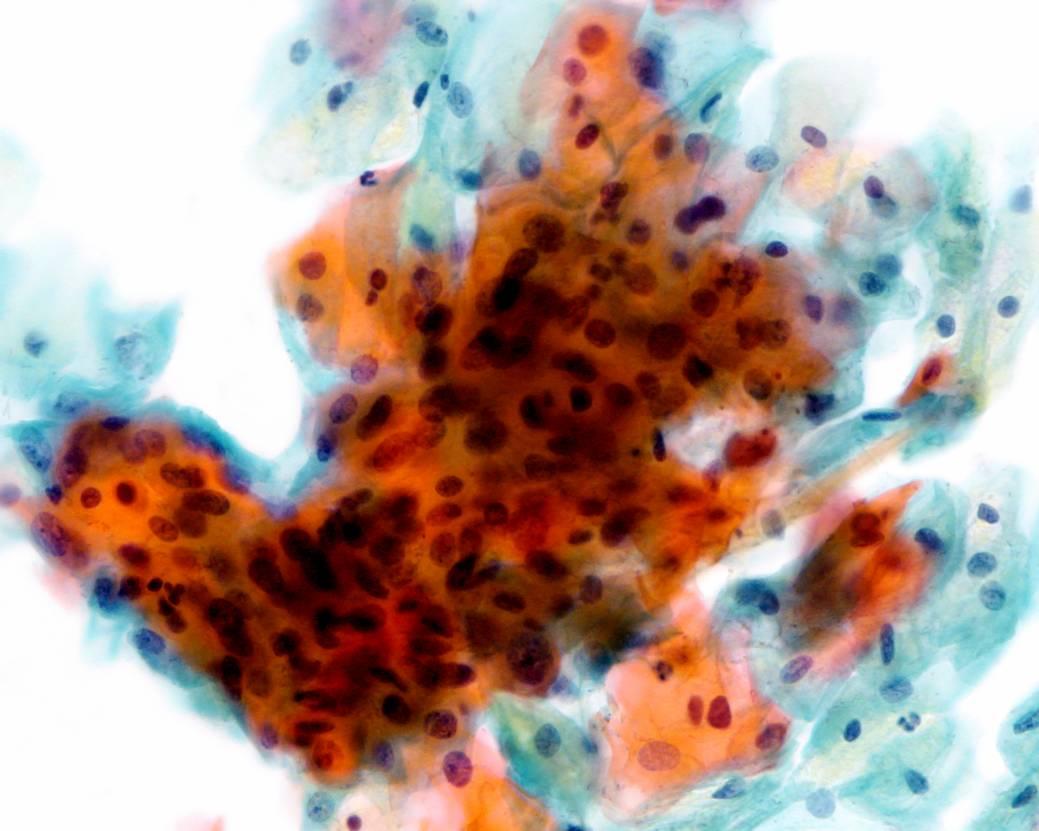
Cytological diagnosis of CIN3 (encompassing HSIL, severe dysplasia and carcinoma in situ)
The Bethesda System definition CIN 3 (HSIL)
- Cells occur singly or grouped in sheets or in syncytial-like clusters
- Abnormal nuclei generally found in cells with immature, delicate or dense cytoplasm; occasionally the cytoplasm is heavily keratinised
- Nuclear enlargement is three or more times the area of normal intermediate nuclei*
- The cytoplasmic area is decreased. For this reason, in a cell with very high nuclear/cytoplasmic ratio, the dimension of the nucleus can be smaller than in LSIL
- Overall cell size is often ksmaller than in LSIL
- Nuclei are hyperchromatic usually with granular coarse chromatin
- Nuclear borders are irregular and anisonucleosis is a prominent feature
- When the abnormal cells occur in sheets ,the nuclei are crowded and overlapping and mitotic figures may be seen
*The WHO cytological definition of CIN 3 requires that the nucleus occupies more than two thirds of the area of the cell.