This content is also available in: English Español

Bookmark (0)

No account yet? Register

Histological section of acute non specific cervicitis showing a the inflammatory response in the stroma characterised by hyperaemia of the papillary vessels, exudation of fluid into the surrounding tissue and migration of polymorphs from the blood stream to the site of the infection. The epithelium is infiltrated  by polymorphs and RBC and the epithelial cells shows  degenerative changes and loss of cohesion. Ultimately the epithelium may be completely ulcerated and the surface epithelium replaced with necrotic debris. 


Cytology of  acute non specific cervicitis

Le caratteristiche dello striscio sono le seguenti:

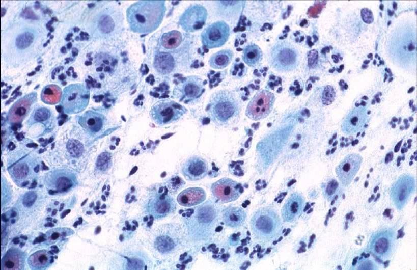
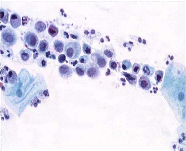
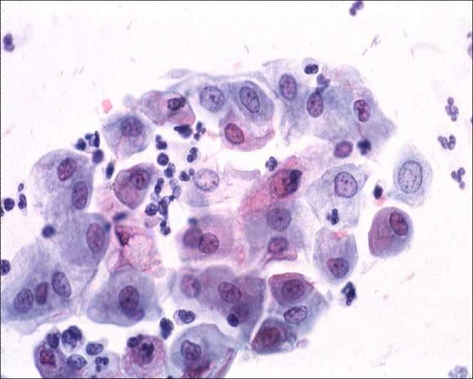
  1. Le cellule epiteliali mostrano marcate alterazioni degenerative a livello citoplasmatico e nucleare. Il citoplasma si mostra vacuolato ed ha un’ aspetto policromatico. Può diminuire in modo cospicuo producendo l’effetto di un alone perinucleare. A volte possono trovarsi polimorfonucleati all’interno del citoplasma. Le modificazioni nucleari variano a seconda dell’ entità del danno. Nelle fasi iniziali mostrano necrosi coagulativa con ammassamento della cromatina, apparente ipercromasia e irregolarità dei contorni della membrana nucleare. Quando si ha una completa lisi cellulare i nuclei diventano rigonfi e sia la membrana che la struttra nucleare diventano indistinte. A volte poi i nuclei assumono un aspetto coartato e sopraggiunge la piccosi o la carioressi.
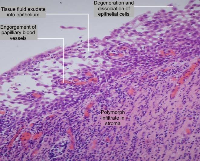
  2. Numerosi polimorfonucleati sono presenti nell’essudato che mostra una alta concentrazione proteica. Occasionalmente i polimorfonucleati sono cosi numerosi che non rendono possibile l’analisi delle cellule epiteliali nello striscio e lo rendono inadeguato per la diagnosi
  3. Emazia, catene di batteri coccoidi o bacilli, detriti cellulari e mucina sono poi presenti come sfondo delle striscio .
Non specific cervicitis: Note degenerative changes in the immature metaplastic cells in the inflammatory smear. The epithelial cells show condensation and clumping of the chromatin in some nuclei and clearing of chromatin in others. The nuclei appear to vary in size. Note vacuolation of the cytoplasm.


Cytology of atrophic cervicitis

Atrophic cervicitis: Atrophic cervices are particularly susceptible to infection. The smears contain numerous polymorphs and mucus strands and the parabasal cells show degenerative changes such as nuclear pyknosis and karyorrhexis. Note polychromatic staining of the cytoplasm.