This content is also available in: Italiano Español Čeština Magyar Polski

Bookmark (0)

No account yet? Register

Inflammatory changes in the cervix

Histological section of acute non specific cervicitis showing a the inflammatory response in the stroma characterised by hyperaemia of the papillary vessels, exudation of fluid into the surrounding tissue and migration of polymorphs from the blood stream to the site of the infection. The epithelium is infiltrated  by polymorphs and RBC and the epithelial cells shows  degenerative changes and loss of cohesion. Ultimately the epithelium may be completely ulcerated and the surface epithelium replaced with necrotic debris. 


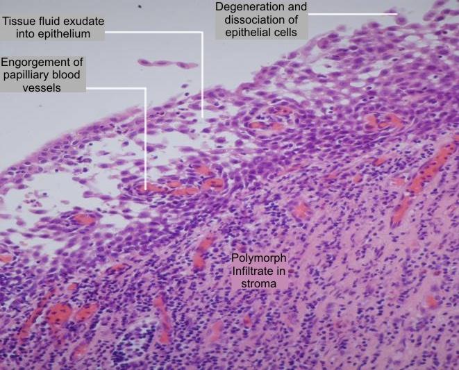
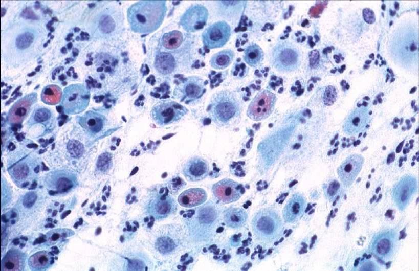
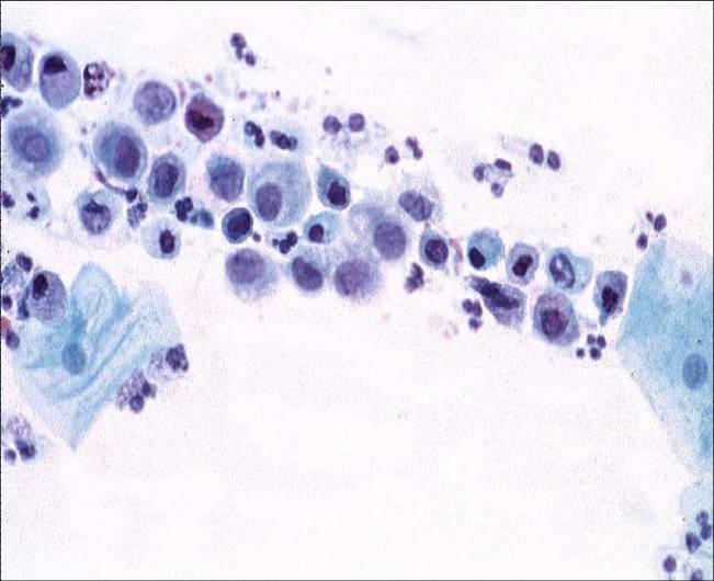
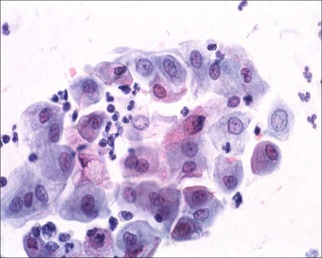
Cytology of  acute non specific cervicitis

The characteristic  features of the smear are:

  1. Epithelial cells which show marked degenerative changes of the cytoplasm and nucleus. The cytoplasm appears frayed or vacuolated and may exhibit polychromatic staining. It may shrink slightly to produce a perinuclear halo. Occasionally polymorphs are engulfed in the cytoplasm. The nuclear changes vary with the severity of the injury. In the earliest stage they show coagulative necrosis with clumping of the chromatin, apparent hyperchromasia and irregularity of the nuclear membrane.  When complete cell lysis supervenes the nuclei become swollen and hypochromatic and the nuclear structure and membrane become indistinct. Eventually the nucleus shrinks and become pyknotic and karyorrhexis supervenes
  2. Numerous polymorphs embedded in a protein rich exudate. Occasionally the polymorphs are so numerous that they obscure the epithelial cells in the smear and render the smear inadequate for evaluation.
  3. Red blood cells, chains of coccoid bacteria or bacilli, mucin and necrotic cell debris may also be seen in the background.
Non specific cervicitis: Note degenerative changes in the immature metaplastic cells in the inflammatory smear. The epithelial cells show condensation and clumping of the chromatin in some nuclei and clearing of chromatin in others. The nuclei appear to vary in size. Note vacuolation of the cytoplasm.


Cytology of atrophic cervicitis

Atrophic cervicitis: Atrophic cervices are particularly susceptible to infection. The smears contain numerous polymorphs and mucus strands and the parabasal cells show degenerative changes such as nuclear pyknosis and karyorrhexis. Note polychromatic staining of the cytoplasm.