This content is also available in:
![]() English
English ![]() Italiano
Italiano
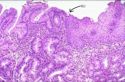
La unión escamocolumnar está localizada en el punto donde el epitelio plano y el epitelio cilíndrico se encuentran. La localización de este punto varía durante la vida de la mujer debido a los cambios metaplásticos en el epitelio cervical que ocurren después de la pubertad y durante el embarazo. La zona de transformación es el nombre asignado al área del cuello uterino compuesta de epitelio que ha sufrido cambio metaplástico.
Definición de metaplasia
-
Metaplasia es el nombre dado al proceso por el cual un tipo de epitelio completamente diferenciado se transforma en otro.
-
Es usualmente un proceso adaptativo que ocurre en reacción a irritación crónica de cualquier clase, o como respuesta a estímulos hormonales.
-
El cambio metaplásico es reversible y teóricamente el epitelio transformado puede revertir al epitelio original después que el estímulo ha desaparecido, pero esto no siempre ocurre.
-
La metaplasia ocurre en muchos sitios del cuerpo p. ej la mucosa gástrica, la vejiga, los bronquios etc. El proceso metaplásico del cuello uterino ha sido estudiado en detalle.
En el cuello uterino, el cambio metaplástico implica la transformación del epitelio endocervical en un epitelio escamoso.
Tres etapas histológicas han sido identificadas:
-
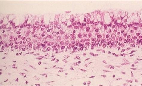
Etapa 1 : Hiperplasia de células de reserva – las células de reserva en el epitelio endocervical empiezan a dividirse.
-
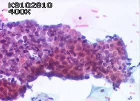
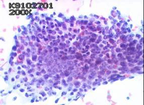
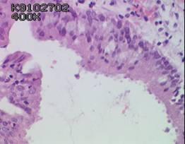
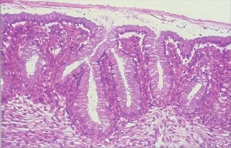
Etapa 2: Metaplasia plana inmadura – las células de reserva proliferan para formar múltiples capas de células indiferenciadas. Una capa superficial de células cilíndricas mucinosas se puede ver frecuentemente en la superficie.
-
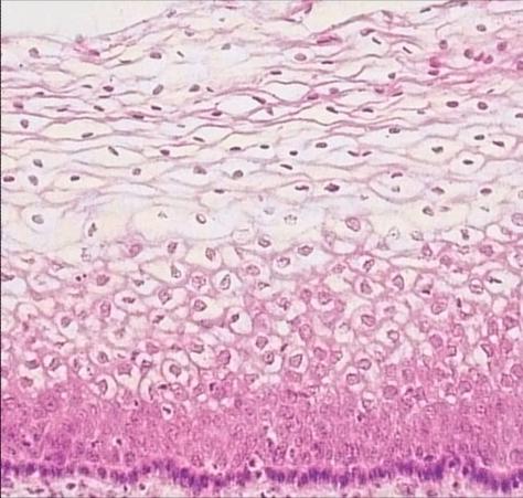
Etapa 3: Metaplasia plana madura – las células indiferenciadas se diferencian en epitelio plano maduro que es casi indistingible del epitelio plano original.
Cambio metaplásico en el cuello uterino y sus bases fisiológicas
-
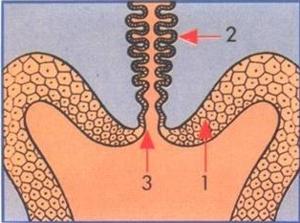
Desde el nacimiento hasta la pubertad, el epitelio endocervical es cilíndrico, y el ectocervix se compone de epitelio plano nativo. La interfase entre estos dos se llama unión escamocolumnar original.
-
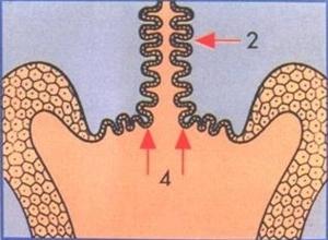
Durante la pubertad y el primer embarazo el cuello uterino aumenta de volumen en respuesta a cambios hormonales. El epitelio endocervical se evierte al ectocervix (portio vaginalis) exponiéndose al pH ácido de la vagina. Esto proporciona un estímulo para el cambio metaplásico del epitelio cilíndrico.
-
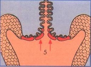
Cambio metaplásico en el cuello uterino y sus bases fisiológicas . Desde el nacimiento hasta la pubertad, el epitelio endocervical es cilíndrico, y el ectocervix se compone de epitelio plano nativo. La interfase entre estos dos se llama unión escamocolumnar original. . Durante la pubertad y el primer embarazo el cuello uterino aumenta de volumen en respuesta a cambios hormonales. El epitelio endocervical se evierte al ectocervix (portio vaginalis) exponiéndose al pH ácido de la vagina. Esto proporciona un estímulo para el cambio metaplásico del epitelio cilíndrico. . El proceso de metaplasia es desigual: comienza en las criptas y en la punta de las vellosidades endocervicales que gradualmente se fusionan. Finalmente la totalidad del epitelio endocervical evertido se reemplaza por epitelio plano.
Noménclatura:
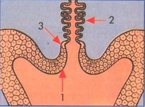
- 1: Epitelio plano nativo
- 2: Epitelio cilíndrico del endocervix
- 3: Unión escamocolumnar (SCJ)
- 4: Eversión del epitelio endocervical
- 5: Cambio metaplástico en la zona de transformación
Significado clínico de la metaplasia plana en el cuello uterino
En el cuello uterino, el área de epitelio que ha sufrido cambio metaplástico se llama la zona de transformación (TZ). Numerosos estudios han demostrado que las células epiteliales metaplásicas inmaduras son susceptibles a carcinogenos, y muchos, si no todos, los cánceres de cuello cervical se original en esta área.
Other forms of metaplastic change in the cervix
Tuboendometriod metaplasia
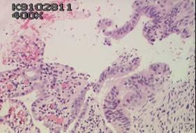
This pattern of metaplastic change often occurs after surgical cone biopsy, diathermy loop biopsy or LLETZ biopsy. Histologically it is characterised by tuboendometrioid glands in the endocervix well away from the uterine isthmus. A key feature of tuboendometrioid change is that there is no associated endometrial stroma The metaplastic epithelium exhibit endometrial features including pseudostratification of the columnar cell nuclei, nuclear hyperchromasia, secretory apical snouting in addition to luminal ciliation.
In cervical smears, tuboendometrioid metaplastic changes may be misinterpreted as glandular neoplasia as the cells may form rather ragged crowded groups. However the absence of feathering and rosette formation and the uniform size and shape and the fine chromatin structure of the individual nuclei should permit the correct diagnosis. The presence of a ciliated cell border is diagnostic although a rare finding. (see also Section on Pitfalls of Diagnosis in Module 10.)
Intestinal metaplasia
This pattern is characterised by the presence of goblet cells in the cervical epithelium and has been found in glandular neoplasms of the cervix.