Tumors
Tumors Benign neoplasms such as urothelial papilloma and inverted papilloma do not have characteristic diagnostic cytological features.
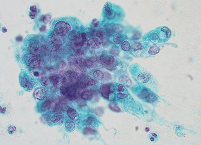
Urothelial carcinoma
Urothelial carcinoma Papillary carcinoma Grade I – voided urine The background is clean Cellularity, consisting almost entirely of single cells, is increased Tumour cells are similar to normal urothelial cells Some loose clusters of urothelial cells The chromatin is vesicular Nucleoli usually absent G1 papillary carcinoma is difficult to diagnose cytologically because of the vesicular […]
Squamous cell carcinoma
Squamous cell carcinoma A squamous component can be seen in cases of poorly differentiated transitional cell carcinoma. In less than 3% of cases it represents the principal part of the tumour. Squamous cell carcinoma is seen most in the regions of the world where Schistosoma haematobium is common. Cytologically the urine contains keratinized malignant squamous […]
Benign conditions
Inflammation Urine is usually turbid The cytological picture shows numerous polymorphonuclear neutrophils and histiocytes. Eosinophils and lymphocytes can also be detected Accentuated reactive changes in epithelial cells Cellular debris Bacteria and other organisms may be present In women it is not uncommon to see Trichomonas vaginalis, Candida albicans and cellular changes induced by polyomavirus (decoy […]
Anatomy and histology
Anatomy of the urinary tract Urine is produced and excreted by the kidneys, two bean-shaped retroperitoneal organs. In the cortical part of the kidney the blood is filtered in the glomeruli. The filtrate passes through the proximal convoluted tubule, the Loop of Henle and the distal convoluted tubule. In these sections of the nephron the […]
Collection of specimens and preparation methods
Collection of specimens Voided urine is the simplest method of collection. Early morning urine should be avoided because of the poor morphological details shown by the cells exfoliated during the night and being exposed to urine for several hours. The best is a mid-morning specimen. The sample should be sent to the laboratory as soon […]
Normal urine
Normal urine Scanty cellularity usually consisting of single cells (especially in voided urine) Umbrella cells and squamous cells seen usually dominate the cytological picture, but deeper layer cells are always seen In catheter samples of urine and in washings clusters or sheets of urothelial cells appear commonly The presence of a few polymorphonuclear neutrophils is […]
Urinary Tract
On completion of this section the cytotechnologist should know: the anatomy of the urinary tract the histology of urothelium the specimen collection methods the preparation techniques the cytology of normal urine cytologic features of inflammatory conditions cytologic features of urothelial carcinoma cytologic features of other types of carcinoma