This content is also available in:
![]() Italiano
Italiano ![]() Español
Español ![]() Čeština
Čeština ![]() Magyar
Magyar ![]() Polski
Polski
Management Guidelines for women with abnormal smears
- International guidelines for the management of women with abnormal smears have been prepared with the support of the Europe Against Cancer program in 1993 (European J of Cancer vol 29A supp 4)
- The guidelines were published with the clear understanding that management decisions are entirely a matter of clinical judgment and beyond the scope of cytology.
- However, some clinicians appreciate management guidance and expect it to be included in the smear report.
- The guidelines presented here were prepared in collaboration with an international team of gynecologists and are based on the current concept of the pathogenesis of cervical cancer and the malignant potential of the various CIN lesions
- It must be remembered that a cytological diagnosis is a presumptive diagnosis. Colposcopy and biopsy are essential in all cases where a cytological diagnosis of neoplasia has been proposed in order to obtain a definitive diagnosis before treatment is started.
- High risk HPV DNA testing is an alternative approach to the management of ASC-US when it can be performed concurrently with repeat cytology. The recommended management for ASC-H (which is a category of reporting used by some cytologist) is colposcopy
| Cytological diagnosis | Management guidance |
|---|---|
| LSIL (incorporating ASCUS and CIN1/mild dysplasia (with and without HPV changes) | Repeat smear in 6 months. If the repeat smear is also reported as LSIL refer for colposcopy. |
| HSIL (incorporating moderate and severe dysplasia, carcinoma in situ CIN2 and CIN3) | Colposcopy advised |
| Suspicious of invasive squamous carcinoma | Colposcopy advised |
| Invasive Squamous carcinoma | Colposcopy advised |
| Endocervical adenocarcinoma in situ ; endocervical adenocarcinoma | Colposcopy advised |
| Atypical glandular cells (endocervical or endometrial or not otherwise specified) | Referral for gynaecological opinion |
| Other malignant lesions | Referral for gynaecological opinion |
Colposcopy
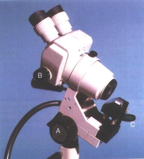
The colposcope is an instrument which permits examination of the uterine cervix, vagina and vulva at high magnification. It consists of a microscope mounted on a mobile stand which has a light source and video camera attached. It was first described by Hinselmann in 1925 and was widely used by gynaecologists as a diagnostic tool for invasive cervical cancer in Germany for many years before it was used to complement cervical cytology for the detection of preinvasive and early invasive cervical cancer.
With the introduction of cervical cytology, the colposcope has become an indispensable tool for the identification and location of lesions in the cervix which give rise to abnormal smear reports. It should be remembered that cervical cancer at its earliest preinvasive stage is rarely detectable to the naked eye and a cytological diagnosis of CIN is at best a presumptive diagnosis. The magnification afforded by the colposcope enlarges the field of view up to x 40 normal size. The magnified images of the cervix enable the colposcopist to do the following:
- Identify abnormal areas in the cervical epithelium and subepithelial angio architecture which are not be visible on normal inspection.
- Precisely locate the lesions and define their limits.
- target areas for biopsy
- carry out histological examination of the biopsy material and obtain a tissue diagnosis
- exclude invasion
- define an appropriate treatment scheme for each woman
- confirm the cytological findings
The process of colposcopy requires that the patient is placed in a modified lithotomy position and the cervix exposed using a bivalve speculum. A repeat cervical smear is often taken at this stage taking care to minimise the risk of bleeding which could subsequently interfere with the field of view. The cervix is then swabbed gently with a cotton swab soaked in saline and inspected. A dilute solution of acetic acid (3% or 5%) is gently applied with the swab and left for approximately five seconds. The acetic acid renders abnormal epithelium visible to the naked eye. Abnormal areas appear as a white usually sharply defined area (aceto white) on the ectocervix. The effect of the dilute acetic acid on the epithelium is believed to be achieved by coagulation of nuclear protein. Because of the high density of nuclear protein in areas of CIN and the changes in angio architecture associated with neoplasia, the areas appear more opaque than the surrounding normal epithelium.
Indications for referral for colposcopy
Before the introduction of colposcopy, women who had abnormal smears or whose cervix looked suspicious were frequently treated by hysterectomy or cone biopsy. Once colposcopy became the routine first step in the investigation of women with abnormal smears, the need for surgical intervention was dictated by the extent, location and type of lesion and the individual preferences of the patient. At present, colposcopy is usually recommended for women who have a smear pattern suggestive of a high grade lesion i.e. CIN2or 3, or CGIN.
Colposcopy is not usually recommended the first time a woman has a smear report suggestive of a low grade lesion (ASCUS or borderline or even LSIL) as these lesions are often transitory. However if a repeat smear indicates that the lesion has persisted for 6 months or more, referral for colposcopy is advisable. Colposcopy is also essential for women who have symptoms or signs of invasive cancer (e.g. intermenstrual or post coital or post menopausal bleeding) even in the presence of a Negative smear report.
Biopsy for the confirmation of cytologic and colposcopic findings
It is worth remembering that neither cytology nor colposcopy are able to provide a definitive diagnosis of neoplastic changes in the cervix. The evidence of CIN from a Pap smear or colposcopic examination of aceto white areas is at best presumptive.
A definitive diagnosis of CIN can only be provided by tissue biopsy and the demonstration of histological changes of CIN in the cervix.
For this reason one or more colposcopically directed punch biopsies should always be taken when an area of aceto white is detected in the cervix. The biopsies should be taken from the most abnormal looking areas of the cervix using a specially designed biopsy forceps with the aim of excluding invasion.
Once a histological diagnosis has been obtained, appropriate treatment can be applied. Ablative techniques such as radical electrocoagulation diathermy, cryocautery, laser vaporisation, and cold coagulation are currently used for the treatment of CIN.
It should be remembered that colposcopic biopsies are generally small and not easy to target so that the most severe pathological changes lesion in the cervix may occasionally be missed on punch biopsy. Large loop excision of the transformation zone (LLETZ) provides a satisfactory solution to this problem as it combines diagnostic biopsy with treatment. The whole lesion is excised by a single sweep of the diathermy loop and all the tissue removed can be processed for histological examination.
Cone biopsy (cold knife or laser) are often reserved for those cases where the lesion appears to be intraepithelial in nature but extend outside the range of colposcopic appraisal or where the cytology suggests a more serious condition than that seen at colposcopy. Cone biopsy is also recommended if the cytology suggests glandular neoplasia as colposcopy can be uninformative in cases of Aden carcinoma in situ or early invasive cervical adenocarcinoma.