This content is also available in:
![]() Italiano
Italiano ![]() Español
Español ![]() Čeština
Čeština ![]() Magyar
Magyar ![]() Polski
Polski
The World Health Organisation (WHO) recognises two main histological types of invasive cancer
- Squamous carcinoma (which constitute about 85% of all cases)
- Adenocarcinoma (which constitute about 10-12% of all cases)
Several other types of carcinoma e.g. adenosquamous carcinoma, adenoid cystic carcinoma, metastatic carcinoma make up the remaining 3-5% of all cases.
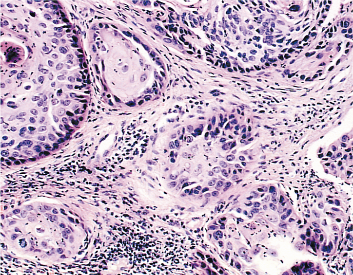
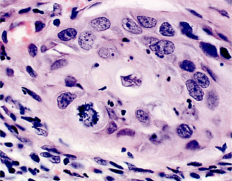
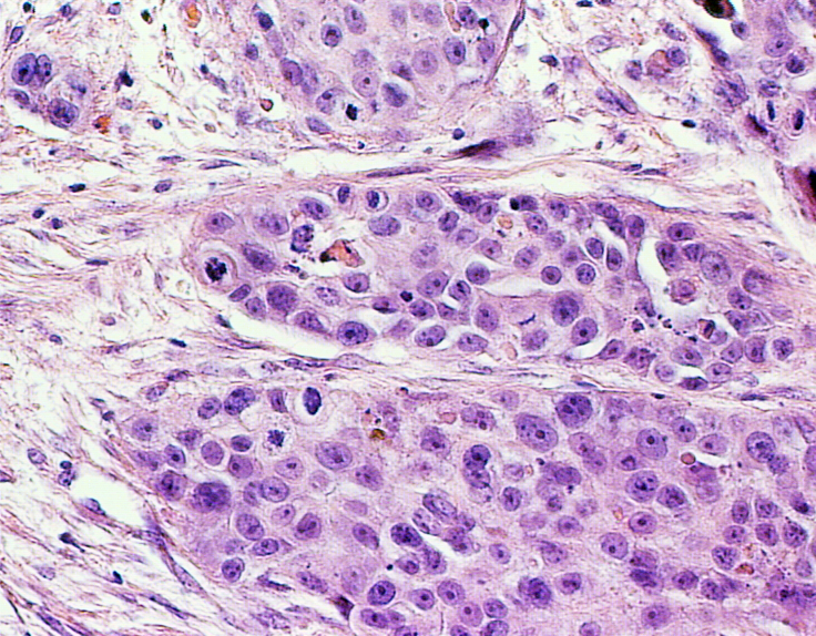
Squamous carcinomas are further typed according to whether they are keratinising or non keratinising carcinomas. Keratinising carcinomas may be well differentiated or moderately differentiated and are composed of large tumour cells. The non keratinising carcinomas (poorly differentiated carcinomas) may be of large cell or small cell type.
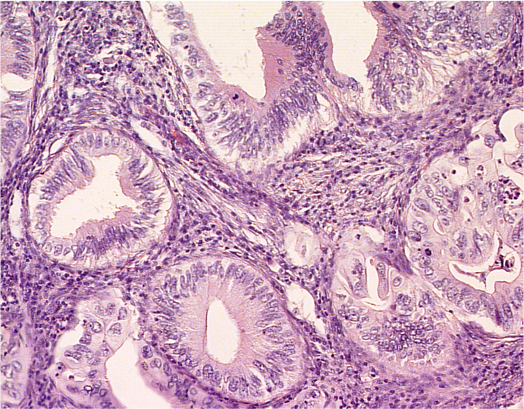
Adenocarcinomas are less commonly found and although each type is histologically distinct it is not uncommon for two or more histological forms of adenocarcinoma to be present in a single tumour. The frequent cooexistence of glandular and squamous carcinoma suggest that they may have a common origin in the reserve cells of the cervix as well as a common aetiology. The most frequent type of adenocarcinoma to be found in the cervix is the endocervical type of mucinous adenocarcinoma. Three grades of endocervical carcinoma are recognised -well differentiated, moderately differentiated and poorly differentiated – depending on the similarity of the tumour cell to the glandular epithelial lining of the endocervix.

Histological types of carcinoma found in cervix (WHO classification)
- Squamous cell carcinoma (epidermoid carcinoma)
- Keratinising (well differentiated and moderately differentiated)
- non keratinising (large and small cell types)
- Spindle cell carcinoma
- Adenocarcinoma endocervical type
- Variant : adenoma malignum (minimal deviation carcinoma)
- Variant :villoglandular papillary adenocarcinoma
- Endometrioid adenocarcinoma
- Clear cell adenocarcinoma
- Serous adenocarcinoma
- Mesonephric adenocarcinoma
- Intestinal type (signet ring) adenocarcinoma
- Other epithelial tumours
- Adenosquamous carcinoma
- Adenoid cystic carcinoma
- Small cell carcinoma
- Undifferentiated carcinoma
- Metastatic tumours (breast, ovary, colon, and direct spread of endometrial carcinoma)