This content is also available in:
![]() Italiano
Italiano ![]() Español
Español ![]() Čeština
Čeština ![]() Magyar
Magyar ![]() Polski
Polski ![]() Türkçe
Türkçe
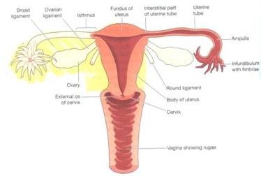
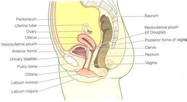
The organs of the female genital tract are located in the pelvis and include the ovaries, the fallopian tubes, the uterus, the cervix and vagina. They are shown in the diagrams below.
The anterior – posterior view shows the relationship of the various parts of the female genital tract to each other. The lateral view shows the relationship of the organs of the female genital tract to the bladder and rectum.
The Ovaries
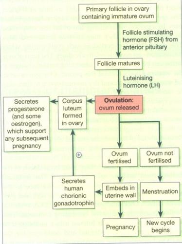
The ovaries are paired organs lying on either side of the uterus adjacent to the lateral wall of the pelvis. They are responsible for oogenesis and the cyclical release of ova in the post pubertal female. The ovaries are endocrine organs producing the hormones oestrogen and progesterone which control the maturation of the ovarian follicles and prepare the uterus for the reception of the fertilised ovum. The production of oestrogen and progesterone is in turn controlled by the gonadotrophic hormones, follicular stimulating hormone (FSH) and luteinising hormone (LH) which are produced by the pituitary gland. FSH and LH regulate the production of oestrogen and progesterone by a feed back mechanism.
The Fallopian tubes
The Fallopian tubes (oviducts) are two fibromuscular tubes about 5 inches (10 cm) long which conduct the ova from the surface of the ovary to the cavity of the uterus. Their funnel shaped ends envelop the surface of the ovary and finger like projections (Fimbriae) direct the ovum into the tube. The ova are conducted along the tubes by a gentle peristaltic movement and by the ciliated epithelium lining the tubes. The epithelium of the Fallopian tubes is thrown into branching folds which provide a suitable environment for fertilisation.